Bacterial Diseases:
Tuberculosis in Poultry It is an infectious disease, typically chronic, affecting humans and domestic animals, caused by Bacillus tuberculosis or its variants, and anatomically characterized by specific cellular vegetations and exudative processes.
But in the beginning, considered identical to the man and mammalian tubercle bacilli of the birds, while other researchers (Maffucci, Strauss and Gamaleia) called attention to the particularities of the bacilli of tuberculosis of the past.
R. Koch cambió, entretanto, de opinión; mas otros investigadores, al frente de ellos Romer y M. Koch y Rabinowitsch (1907), siguieron creyendo que los bacilos de la tuberculosis de las aves eran idénticos a los de la tuberculosis del hombre y mamíferos.
Since they had managed to infect experimentally hens with material sick of people and of animals, mammals and isolate in pure culture for acid-fast bacilli of tuberculosis, avian alterations-tuberculous mycobacteria.
This idea was later abandoned, due to the fact, that in hens exempt from all tuberculosis infection, as evidenced by the test tuberculínica, the experimental infection by tubercle bacilli of human or bovine occurred only in the worst case, alterations, local cured, but never injury-tuberculous mycobacteria such as those presented in the tuberculosis spontaneous fowls (Tschebnitz 1923, Richter, R. Eber, and others).
It was found, furthermore, that the bacilli of tuberculosis of birds may produce alterations of tuberculosis in pigs (O. Bang, Mohler and Washburn), and, in isolated cases, in other mammals.
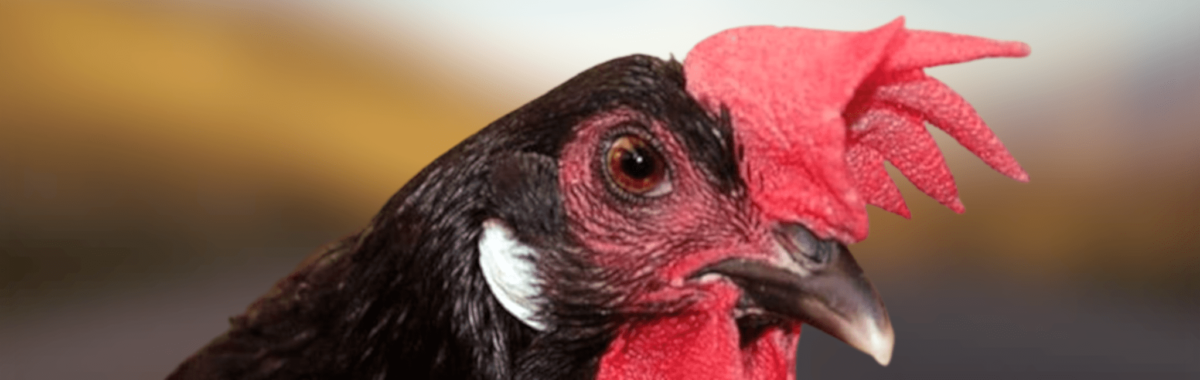
Tuberculosis of the poultry occurs with some frequency and cause sensitive losses in poultry houses and dovecotes. It is also observed in other avian (turkeys, guinea fowl); instead, it is exceptional in waterfowl.
Exotic birds, such as parrots, canaries, and similar species, are also frequently affected. It is also occasionally observed in wild birds (partridges, pheasants, capercaillies, grouse, choughs, etc.).
The apparent recent increase in the incidence of avian tuberculosis appears to be linked to the poor housing and hygiene conditions in which many poultry are kept in cities. However, the disease is not uncommon on small rural farms either, where it likely spreads widely because the birds are often slaughtered only at an advanced age.
In contrast, in large-scale farms poultry the degree of infection decreases from that, lately, they tend to sacrifice the breeding birds in the meet 2 years of age and will keep the pups away from the adult birds.
Tuberculosis in poultry is caused by the avian tuberculosis bacillus (the gallinaceous type of the tuberculosis bacillus). Only in pet birds is tuberculosis most often caused by human tuberculosis bacilli.
Infections occasional poultry by bacilli of tuberculosis, human or bovine, even when operating, lack of practical importance, since at most produce alterations in local and prone to healing.
Usually occurs by ingestion of organs of tuberculosis or deletions of sick birds. It is also produced by wounds of the oral mucosa and of the skin, and it is not impossible to infection by the respiratory tract.
The infectious agent is usually introduced into healthy poultry houses by poultry infected with tuberculosis or by material contaminated with bacilli-containing excretions from such animals.
Tuberculosis bacilli are found relatively frequently in the eggs of tuberculous chickens (according to Fitch, Lubbe, Husen, and Dykmans 1928, up to 1% of eggs from chickens that simply react to tuberculin should be considered infected). Ten days after infection, the bacilli may already have settled in the ovary so that, from this point on, the hens usually lay infected eggs (Raebiger 1929).
Despite this, the infection germination would have no great importance, since the eggs or infected do not develop embryos (Fitch and Lubbe Husen 1924, F. Schmidt) or, if they develop, the chicks die immediately (Baumgarten). In practice, the infection by eggs that contain bacilli can only happen when you feed the birds with raw eggs.
Según Scheible (1950) y otros, en la mayor parte de los casos pueden encontrarse bacilos también en la sangre y en la carne de las aves muertas o sacrificadas a causa de su reacción positiva a la prueba de tuberculina, los cuales pueden ser motivo para el contagio de gallinas.
In the pens infected, the chances of contagion, and the number of cases depend on of the conditions in which they have birds, as the lack of cleanliness and the crowding of the averío in local small and little care favor infection.
The age at which birds suffer from tuberculosis also depends on local conditions. In large breeding farms, where chicks are isolated from adult birds for some time, cases of tuberculosis do not usually occur in birds less than 1 year old; however, in small chicken coops where chicks are in contact with adult birds with tuberculosis from the outset, the disease is already observed in young birds.
Even on small farms, the number of cases of illness and death increases with age.
Poor hygiene conditions and, above all, inadequate nutrition influence the course of the disease, since in well-maintained pens, weight loss in birds due to tuberculosis is usually observed at a later age than in poorly fed flocks.
Once tuberculosis bacilli reach the digestive tract, they penetrate through the lymphatic follicles of the pharynx or intestine into the deeper layers of the tissues, regardless of whether or not they have caused pathological changes in the digestive tract.
They enter the bloodstream and are carried by the blood to various organs, particularly the spleen, liver, bones, etc., where they cause tuberculous lesions.
Tuberculosis in birds always begins with bacteremia, which, in severe infections, can occur as early as 2 to 3 days later (Raebiger).
As the disease progresses, the bacilli from the foci formed in the manner described may temporarily enter the bloodstream and reach organs that have not yet been affected. In addition, tuberculosis bacilli reach the liver not only via the bloodstream but also through the lymphatic system.
In the rare cases in which the infection occurs through the respiratory tract or cutaneous wounds, the primary focus develops in the lungs or on the skin, where it then the bacilli enter also in the way blood count and go with the blood to the organs more diverse.
The demonstrations caquécticas, which ensue in advanced cases, are the result of the toxic action of the chemicals of the bacillus and the tissues destroyed. The death ensues, he prays as a result of cachexia, prays for internal bleeding, particularly of the liver and spleen.
Anatomical alterations:
In the necropsy is sick most often the liver, which is increased volume and color, as red, brown, already noticeably yellow or gray-yellow. In advanced cases, presents degeneration of adipose and is brittle, friable. The number and size of foci of tubercular that contains vary a lot.
Small spots the size of a millet seed (miliary) and even smaller ones (submiliary) are glass-clear, grayish-white, or white, and sometimes yellowish as well.
On sectioning, they appear uniformly hyaline or with a small yellowish spot in the center. While the small lesions do not protrude from the surface of the liver, the larger ones form more or less prominent nodules. Lesions that reach the size of peas are yellow or yellowish-brown, and their surface may be transparent or opaque.
The cross-section of such lesions is hyaline at the periphery and has a hard-cheese-like appearance in the center; more rarely, the entire surface is soft, or only the central part is soft.
The even higher and can reach the size of hazelnuts and even nuts.
They now resemble spheres, especially when they result from the fusion of several nodules; their surface resembles that of cauliflower. The larger nodules are yellow and hard as cartilage or even harder, so that they can be cut only with difficulty, but when cut they do not usually crunch, because they contain calcium salts only in rare cases.
Their cross-sectional surface is also yellow or yellowish-gray, but rarely uniform. They are characterized by concentric rings resembling the pattern of agate, or by light-yellow or dark-yellow spots mixed with dull gray ones, so that the cross-sectional surface resembles the polished surface of marble conglomerates.
Because the tuberculous tissue of the liver is extremely fragile, hemorrhages beneath Glisson’s capsule are common, as is (according to A. Eber, in approximately 20% of cases) rupture of the capsule; consequently, in such cases, signs of internal bleeding are found alongside liver tuberculosis.
The spleen is also injured often. Generally increased volume, the times to be that of a hen's egg.
The spotlights are similar to those of the liver, but tend to be smaller. Exceptionally, you see a few very large, and until all the organ can be found transformed into a mass cheesy.
In some cases, it is also observed hemorrhages under the capsule breaks and the same with internal bleeding.
Symptom:
Usually, it takes months to manifest phenomena morbid. In some cases, a limp singular, of a leg, wake up early suspicion of tuberculosis of bone, but, more often than not, only that they wake up the emaciation progressive, despite maintaining the appetite and can even be increased, and anemia.
Later ensues loss of appetite, decreases or stops, all the laying of the eggs, and the birds that have been left in the meantime on the bones, they die after a period of 6 to 8 weeks of overt illness, if before the break of any organ tuberculosis does not put an end to life due to internal bleeding.
In addition to these signs morbid, vague, with time elevation of the temperature, in other cases it may be useful for the diagnosis of phenomena that follow.
The intestinal tuberculosis is manifested by diarrhea, irrepressible and, in birds, very gaunt, through the abdominal wall can be seen on the tubers of the intestine.
Sometimes tuberculous nodules can also be felt in the liver (Jakob suggested examining the abdominal organs after opening the abdomen—for example, during castration—but this is a time-consuming procedure and is not usually performed).
Tuberculosis of the glands of the neck is manifested by a swelling the size of a hazelnut that can't slip into the stomach, and this distinguishes it from kernels of corn stuck.
In tuberculosis of the joints (in the carrier pigeon is, with the paratifus, the most frequent cause of paralysis of the wing), there are, in addition to pain and swelling of the joints, motor disorders (claudication, fall of the wing a sick, inability to fly).
Sometimes the lump joint is opened, flowing through the open hole a secretion cheesy.
In cutaneous tuberculosis, small nodules—either isolated or in clusters—often develop around the eyes, on the skin, and in the subcutaneous tissue; these gradually grow to the size of peas. Later, the larger nodules rupture and develop into ulcers with eroded edges and sluggish granulation tissue covered with caseous material.
In a case of Pallaske (1942), the fund ulceroso, covered in pus and scabs, had a look sarcoid, without caseificación, which histologically is characterized by a proliferation of epithelial cells giants. In a case of abscess of the pulpejos, Hepping (1938) found tubercle bacilli in the content evacuated prior to incision.
In the living animal, usually only allow you to diagnose the disease, the demonstration of tubercle bacilli in the deletions and secretions or the positive result of the test tuberculínica, by the great similarity of the clinical manifestations with those of other diseases (verminosis, cholera in poultry, typhus of the same, paratifus).
In the post-mortem examination is usually sufficient to diagnose the finding of foci circumscribed, consistent, often, hardness cartilaginous, with caseificación or softening plants.
In these cases deserve special attention in the bone alterations, which affect, in the majority of cases of tuberculosis, the long bones already mentioned.
In cases of uncertainty, particularly when lesions are very recent, they may closely resemble those of other infectious diseases (poultry typhus, paratyphoid, pseudotuberculosis, aspergillosis); in addition to a necropsy, microscopic examination of the affected tissues is necessary.
To discover tuberculous infection in poultry, the intradermal test of Van Es and Schalk (1914) is adequate; in contrast, the subcutaneous tuberculin and ocular tuberculin tests are inadequate.
A positive intradermal test result is a certain sign of tuberculous infection; a negative result indicates with less certainty the absence of tuberculosis.
Often, the positive reaction is not present in birds very fall, precisely because of tuberculosis, and, on the other hand, takes up to three weeks after infection, before the animal will react to the tuberculin.
For this reason, in emaciated birds, only the positive result of the tuberculin test is of value and, even in robust birds, the negative result only decides the exclusion of tuberculous infection when the test, repeated at least three weeks later, is negative again.
Techniques and interpretation of the test tuberculin:
In the chicken, is injected in the middle of the anterior edge of the chin to 0.1 c. c. approximately tuberculin poultry or concentrated to 50 % in the skin (in case of need, also can be injected into an appendage or on the crest).
If the test is to be repeated, the other chin should be used, since, according to Revesz (1931), tissue that has been injected once with tuberculin remains unresponsive to it for a period ranging from a few weeks to two months (a finding not confirmed by Bamberger (1942)).
Under practical conditions, non-tuberculous birds are not rendered sensitive to tuberculin by injections of the latter (according to Radtke, at least seven injections are necessary for this, carried out at intervals of one week).
Shortly after the injection, the area surrounding the point injected swell even in birds are not infected, because of the injury of the tissues and the contents of irritating non-specific contained in the tuberculin.
In uninfected birds, this specific swelling disappears within 24 to 36 hours; in contrast, in chickens that react positively, the swelling develops into a specific swelling that is very evident after 36 to 48 hours and gradually subsides, sometimes taking 7 to 8 days. For this reason, the test should be evaluated 36 to 48 hours after the injection.
Positive reaction is manifested by a consistent, circumscribed swelling in the immediate vicinity of the injected spot. If measured with a small caliper, increases in thickness of more than 2 mm can be regarded as signs of positive reaction.
Can be confused very easily with the reaction tuberculínica positive, solid masses that are observed, in some chicken coops, after the intradermal injection of tuberculin, in birds carrying bacilli of cholera in chickens.
Because Pasteurella bacteria, carried to the tuberculin injection site by the blood, attach themselves there and, by multiplying in the damaged tissue, can cause chin disease.
But in that case, the swelling is edematous and spreads across the entire chin and, sometimes, also to the throat and the other side of the chin, and it usually continues to spread even 48 hours later.
Since Moses, Feldmann and Mann (1943) found the agglutination test satisfactory in laboratory experiments, using strains of avian tuberculosis bacilli that emulsified well, for the examination of serum from chickens suspected of having tuberculosis, Larson and Feloman (1950) developed a slide-based method applicable to the hematological examination of birds in the henhouse.
This fresh-blood method has rendered tuberculin tests unnecessary and can also be used to detect typhoid infections in birds. The results obtained to date are encouraging, although a definitive assessment of the method’s effectiveness is still pending.
Tuberculosis of poultry is considered today as an incurable disease, for which the fight against the disease should be established on the basis of the slaughter of sick animals and to preserve the health of those not infected yet.
The pens are not infected should not be, but birds from farms free of tuberculosis (in this sense, it is of importance, the official recognition of such farms, as exempt from tuberculosis) and, if this is not possible, the acquired again to remain isolated and will be submitted twice to the test tuberculin with the interval of 3 to 4 weeks, depending on your employment as a breeding bird of the good result of the research.
Once the presence of tuberculosis has been confirmed in a flock, not only the visibly sick birds but also those that merely react to the tuberculin test must be culled; the healthy portion of the flock should be moved to a tuberculosis-free facility.
By means of a new investigation, carried out 3 to 4 weeks later (and, later, at least annually), and the elimination of the reacting birds, the pen is kept definitively free of tuberculosis.
Bibliography:
Anderson, Miller DA, Trampel DW. Epidemiological investigation, cleanup, and eradication of pullorum disease in adult chickens and ducks in two small-farm flocks. Avian Dis. 2006; 50(1):142-7.
Basnet HB, Kwon HJ, Cho SH, Kim SJ, Yoo HS, Park YH, Yoon SI, Shin NS, Youn HJ. Reproduction of fowl typhoid by respiratory challenge with Salmonella Gallinarum. Avian Dis. 2008; 52(1):156-9.
Berchieri Jr., Murphy CK, Marston K, Barrow PA. Observations on the persistence and vertical transmission of Salmonella enterica serovars Pullorum and Gallinarum in chickens: effect of bacterial and host genetic background. Avian Pathol. 2001; 30(3):221-31.
Cobb SP, McVicar CM, Davies RH, Ainsworth H. Fowl typhoid in caged layer birds. Vet Rec. 2005; 157(9):268.
Kahn CM, Line S, editors. The Merck veterinary manual [online]. Whitehouse Station, NJ: Merck and Co., 2006. Fowl typhoid. Available at: http://www.merckvetmanual.com/mvm/index.jsp?cfile=htm/bc/204103.htm. Accessed 16 Jun 2009.
Lister, S. A., & Barrow, P. Fowl typhoid (Salmonella Gallinarum infection). In: Pattison M, McMullin PF, Bradbury JM, Alexander DJ, editors. Poultry diseases. 6th ed. Philadelphia, PA: Saunders Elsevier; 2008. p. 130-3.
Lister, SA, and Barrow, P. Pullorum disease. In: Pattison M, McMullin PF, Bradbury JM, Alexander DJ, editors. Poultry diseases. 6th ed. Philadelphia, PA: Saunders Elsevier; 2008. p. 126-30.
Mississippi State University Cooperative Extension Service. Bacterial diseases: fowl typhoid [online]. Mississippi State University; 1997. Available at: http://www.msstate.edu/dept/poultry/disbact.htm. * Accessed 11 Oct 2001.
Pennycott, T. Diseases of game birds: Gives. In: Pattison, M, McMullin PF, Bradbury JM, Alexander DJ, editors. Poultry diseases. 6th ed. Philadelphia, PA: Saunders Elsevier; 2008. p. 562-3.
Pennycott TW, Duncan G. Salmonella pullorum in the common pheasant (Phasianus colchicus). Vet Rec. 1999; 144(11):283-7.
Shivaprasad HL, Barrow PA. Pullorum disease and fowl typhoid. In: Saif YM, Fadley AM, Glisson JR, McDougald LR, Nolan LK, Swayne DE, editors. Diseases of poultry. 12th ed. Ames, IA: Blackwell Publishing; 2008. p. 620-34.
Shivaprasad HL. Fowl typhoid and pullorum disease. Rev Sci Tech. 2000; 19(2):405-24.
World Organization for Animal Health (OIE). World animal health information database (WAHID) [database online]. Fowl typhoid. Paris: OIE; 2009 Mar. Available at: http://www.oie.int/wahid-prod/public.php. Accessed 16 Jun 2009.
World Organization for Animal Health (OIE). World animal health information database (WAHID) [database online]. pullorum disease: Paris: OIE; 2009 Mar. Available at: http://www.oie.int/wahid-prod/public.php. Accessed 16 Jun 2009.
World Organization for Animal Health [OIE]. Manual of diagnostic tests and vaccines for terrestrial animals [online]. París: OIE; 2008. Fowl typhoid and pullorum disease. Available at: http://www.oie.int/eng/normes/mmanual/2008/pdf/2.03.11_FOWL_TYPHOID.pdf. Accessed 16 Jun 2009.
Internet resources:
Food and Agriculture Organization of the United Nations (FAO). Manual for the Recognition of Exotic Diseases of Livestock
The Merck Veterinary Manual http://www.merckvetmanual.com/mvm/index.jsp
World Organization for Animal Health (OIE) http://www.oie.int/es/
OIE Manual of Diagnostic Tests and Vaccines for Terrestrial Animals
You may be interested in these other sections