- Home
- Contact TRI-TRO
- Discovering the Ancestors of the Poultry Spanish
- Cock Spanish Black: features, origin and behavior
- The Origin of the Domestic Chicken: Gallus and the Evolution of the Species
- Castilian Black Chicken
- Practical Poultry Farming
- Anatomy of the Rooster
- Selection in Poultry Farming: Genetic Improvement and Breeding
- External morphology of the hen: parts and structure
- Anatomy of the chicken: parts, organs, and functions
- Castilian Black Hen: an elegant, egg-laying Spanish breed
- The pecking order: social hierarchy among chickens and roosters
- Chicken Droppings: A Visual Guide to Interpreting Their Excrement
- Broody hens: a step-by-step guide to natural incubation
- The chicken egg: how it forms and its parts
- Chick Sexing: How to Tell if a Chick Is Male or Female (1)
- Castilian Black Chicks: Initial Care
- Incubating Chicken Eggs: A Complete Guide
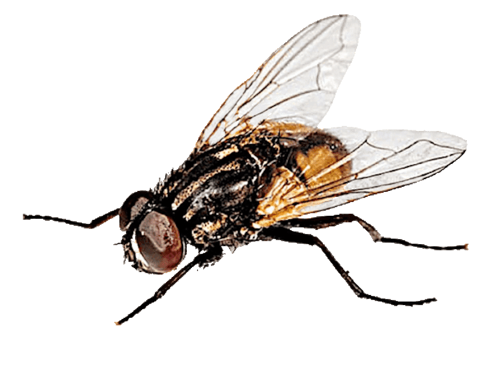
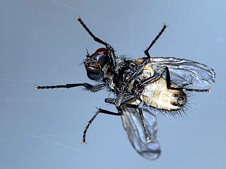
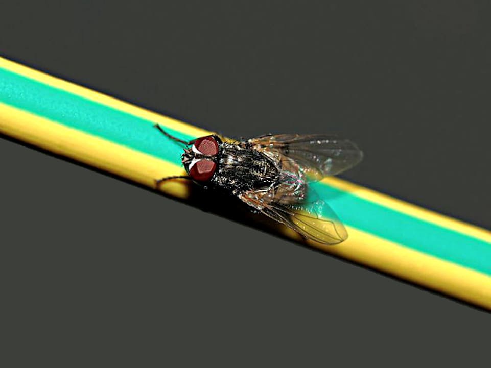
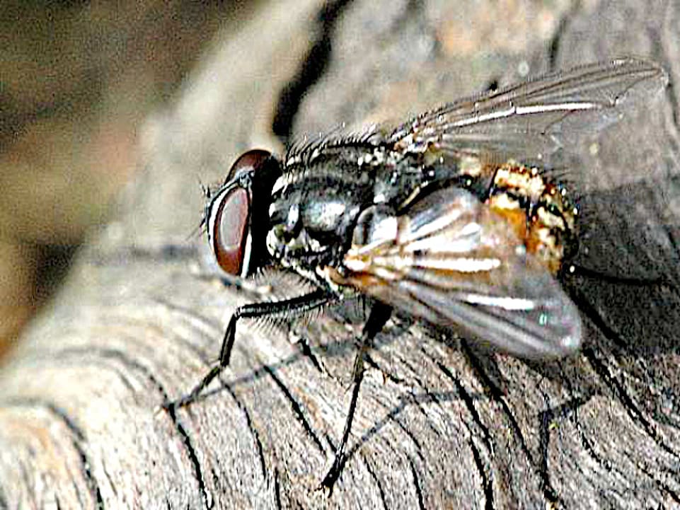
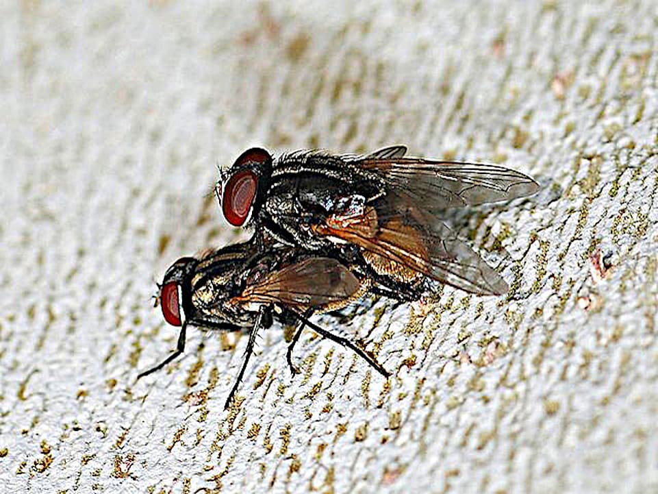
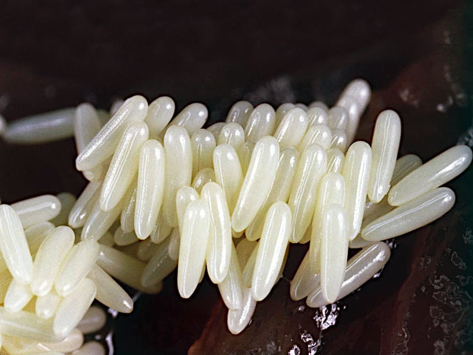
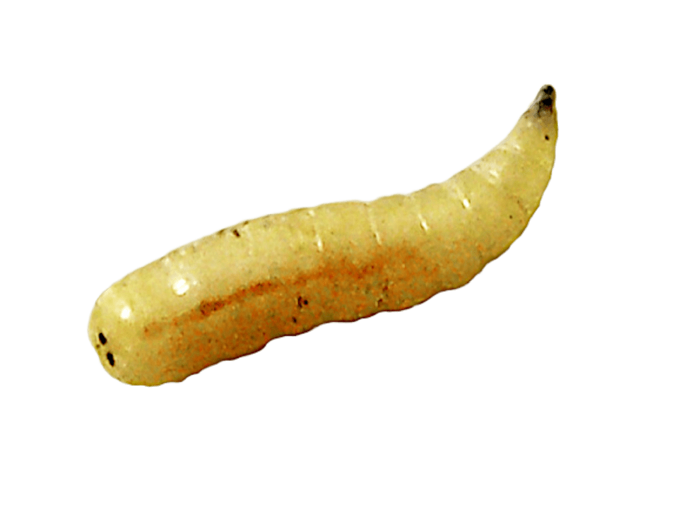
- Flies in the chicken coop and how they affect the chickens
- Chicken Diseases: A Comprehensive Guide, Symptoms, and Types
- Bacterial Diseases in Chickens: Symptoms, Transmission, and Prevention
- Salmonella in chickens: symptoms, transmission, and treatment
- Pullorosis in hens: white bacillary diarrhea and treatment
- Infectious coryza in chickens: symptoms and treatment
- Colibacillosis in chickens: symptoms and treatment
- CRD in hens: respiratory disease and treatment
- Avian cholera in chickens: symptoms and treatment
- Infectious synovitis in hens: symptoms and treatment
- Swollen Head Syndrome in Chickens (SICH): Symptoms and Treatment
- Other bacterial diseases in chickens
- Avian typhus in chickens: symptoms and treatment
- Omphalitis in chicks: navel infection and treatment
- Necrotic enteritis in hens: symptoms and treatment
- Avian Tuberculosis in Hens: Symptoms and Transmission
- Staphylococcal infection in chickens: symptoms and treatment
- Streptococcus in chickens: symptoms and treatment
- Avian erysipelas in chickens (red disease): symptoms and treatment
- Pseudomoniasis in chickens: symptoms and treatment
- Spirochetosis in chickens: symptoms and treatment
- Avian Listeriosis in Chickens: Symptoms and Treatment
- Klebsiella in chickens: bacterial infection and treatment
- Viral Diseases in Chickens: Symptoms, Transmission, and Prevention
- Avian Influenza in Chickens: Symptoms, Transmission, and Prevention
- Newcastle Disease in Chickens: Symptoms and Prevention
- Marek's Disease in Chickens: Symptoms and Treatment
- Infectious Bronchitis in Chickens: Symptoms and Treatment
- Gumboro Disease in Chickens: Symptoms and Prevention
- Infectious laryngotracheitis in chickens: symptoms and treatment
- Egg-Laying Drop Syndrome in Chickens: Symptoms and Prevention
- Fowlpox in Chickens: Symptoms and Treatment
- Other viral diseases in chickens
- Parasitic Diseases in Chickens: Symptoms and How to Treat Them
- Red Mites in Chickens: Symptoms and Treatment
- Lice in chickens: symptoms and how to get rid of them
- Coccidiosis in Chicks and Hens: Symptoms and Treatment
- Scabies in Chickens: Causes and Treatment
- Cecal ascariasis in chickens: symptoms and treatment
- Fleas on chickens: symptoms and control
- Chicken Ticks: Symptoms and Treatment
- Capillariosis in Hens: Symptoms and Treatment
- Other parasitic diseases in chickens
- Gizzard worms in chickens: symptoms and treatment
- Intestinal Ascaridiasis in Chickens: Symptoms and Treatment
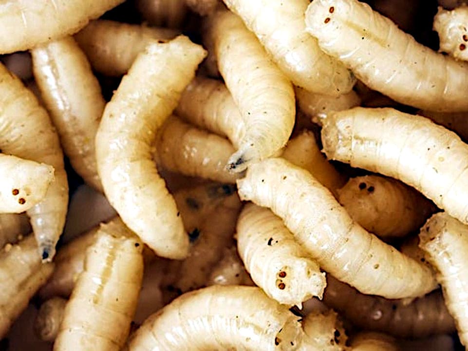
- Chicken Maggot: Control and Risks
- Hexamitiasis in chickens: symptoms and treatment
- Mosquitoes in chicken coops and transmitted diseases
- Mange in Laying Chickens: Symptoms and Treatment
- Scaly Mange in Chickens: Symptoms and Treatment
- Air Sac Mange in Chickens: Symptoms and Treatment
- Syngamus trachea in chickens: symptoms and treatment
- Tapeworm Infection in Chickens: Symptoms and Treatment
- Tetramerosis in hens: symptoms and treatment
- Toxoplasmosis in chickens: causes and prevention.
- Genital trematodiasis in chickens: symptoms and treatment
- Trichomoniasis in chickens: symptoms and treatment
- Sarcosporidiosis in chickens: symptoms and treatment
- Fungal Diseases in Chickens: Types, Symptoms, and Treatment
- Other common health problems in chickens
- Cloacitis in chickens: causes, symptoms, and treatment
- Bruises and contusions in chickens
- Hip and back injuries in hens
- Wounds in chickens
- Stress and anxiety in chickens
- Gizzard inflammation in chickens (inflammation of the gizzard)
- Pecking or cannibalism in chickens
- Oviduct prolapse in chickens
- Abdominal egg-laying in chickens
- Ascites in hens: swollen abdomen
- Chest lumps or blisters in chickens
- Bacterial Diseases in Chickens: Symptoms, Transmission, and Prevention
- Hens. Spanish poultry breeds.
- How I made my chicken coops and more
- Feeding the chickens
- Vitamins for our chickens
- Types of Crests, Combs on Cocks
- Videos Castilian hen
- Hens of the World Prints
- Our Hens: News
- Links Aviculture
- GANECA
- VII Championship of Spain. Alba de Tormes 2012
- International exhibition of Poultry – The Pinina Vegadeo 2012
- Photos Castilian Vegadeo 2012
- Castilian Hen, 7th Spanish Championship