Epidemiology:
The life cycle of Capillaria contorta, C. obsignata and C. anatis It is direct. The parasite's eggs are shed in the feces and develop in the external environment; the larva remains inside the egg and becomes infectious within 2–3 weeks.
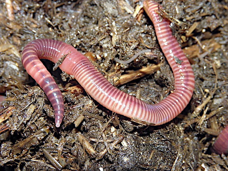
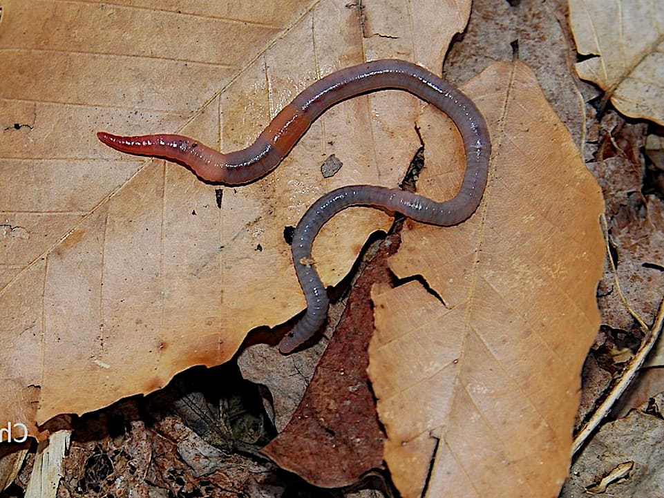
El hospedador se infecta cuando ingiere los huevos al picotear en el suelo. Sin embargo, en el caso de C. contorta, se admite que las earthworms (Eisenia fetida and Allolobophora caliginosa) may act as carriers of infective eggs, and it is even possible that the cycle could be direct or indirect, with earthworms serving as true intermediate hosts.
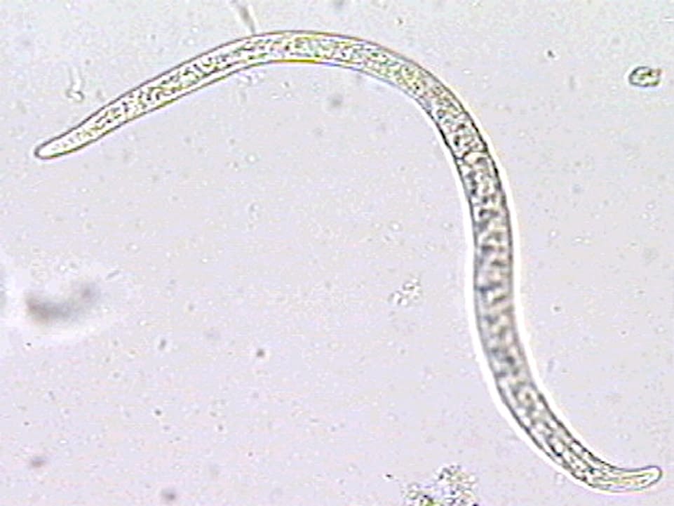
Eggs excreted in feces develop into first-stage larvae in the environment within 11–12 days. Earthworms ingest the larval eggs, and the larvae reach the infective stage within the worms about 9 days after ingestion, once they have shed their eggshells in the earthworms’ digestive tract. Once birds become infected by ingesting the earthworms, the worms develop and reach the adult stage in about 3 weeks.
Pathogenesis:
A small number of worms of any species do not cause problems in birds. On the other hand, if they are very numerous, they cause severe clinical symptoms that affect the economic performance of infected birds and can lead to their death.
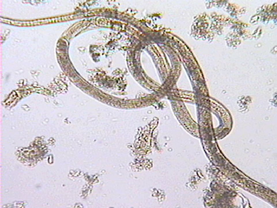
The larvae and adult worms penetrate the mucosa of the esophagus and crop, or bowel.
As a result, the mucosa is inflamed, the higher the number of parasites.
The epithelium sloughs off, and hemorrhagic spots appear on the mucosa. The walls of the esophagus and crop thicken, and their musculature loses tone; the mucosa becomes coated with copious mucus, and ingested food remains trapped in the crop or is emptied slowly into the proventriculus.
If the small intestine is affected, enteritis develops, initially affecting the mucosa; later, pinpoint hemorrhages appear and the epithelium sloughs off, secretions increase, and intestinal absorption decreases, leading to watery diarrhea.
If the parasites are found in the ceca, the mucous membrane becomes inflamed, leading to diarrhea, which is usually very thick and sticky. In all cases, the birds’ overall health and productivity suffer; they lose weight and may even die.
Symptoms:
Capillariasis is observed in birds of all ages, and adult birds act as carriers and spreaders of the worms.
Symptoms appear as early as the pre-clinical stage. In cases of intestinal Capillaria infections, animals experience weight loss, diarrhea with pasty, sticky, and foul-smelling stools, poor general condition, loss of appetite, and reduced water intake.
The birds spend a lot of time huddled on the ground, with their eyes closed, their necks bent, and their heads resting on their crops. Their feathers are ruffled and mottled around the vent, and their visible skin and mucous membranes are pale. Some birds lose weight, become extremely thin, and die.
In infections by species occurring in the esophagus and crop, the birds are downs, weak and thin. Move only when they are annoyed and groggy. Sometimes adopt the posture of a penguin, resting on the feet.
Extend and retract their heads and necks as in an attempt to swallow it, or remove an obstruction.
Injury:
A small number of Capillaria worms do not cause lesions, but a significant number lead to inflammation of the mucosa of varying severity, ranging from catarrhal to croupous (cough), with the formation of pseudodiphtheritic membranes.
In infections by C. contorta and C. annulata are thickened the walls of the esophagus and crop, with the mucous covered with a swab or granulated coposo and, especially in turkeys, the crop can contain a liquid with a foul odor, which is paused on that body by the obstruction of the light due to the thickening of the walls.
With the species C. caudinflata and C. obsignata, the small intestine is observed inflamed and mucosal destroyed and the intestinal lumen, more or less blocked by the scarring of mucous loosened. The mucosa may present petechiae or diffuse redness. The blind affected by C. anatis have lesions similar to those caused by other species in the small intestine, and the contents of the blind is strongly viscous.
Diagnosis:
Is set usually at autopsy, examining the bodies washed in the identification of the parasites. Through coprología can be discovered eggs of characteristic form, and, by counts, you can judge the importance of the parasitación.
Prognosis:
Should be judged taking into account the importance of the infection in the birds they support. In any case, infections, moderate or intense, can lead to serious economic repercussions.