Parasitic diseases:
Sarcocystosis is a disease caused by protozoa, which is distinguished by generate alterations in the striated muscles of the birds and other animal species.
In the case of birds, the causative agent is Sarcocystis horvathi. Se presenta como una infección lenta y sigilosa que afecta predominantemente a las aves adultas. Sarcocystis, término que proviene de las palabras griegas sarx (carne) y kystis (vejiga), se clasifica como protozoos del grupo Apicomplexa, que se desplazan y penetran las células, provocando sarcosporidiasis o sarcocistiosis.
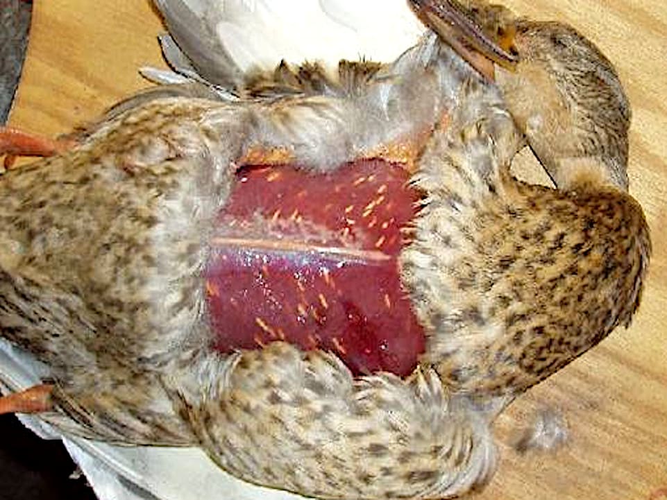
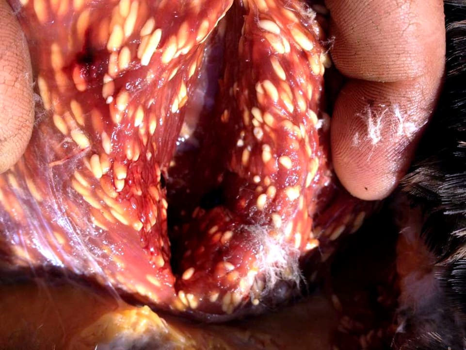
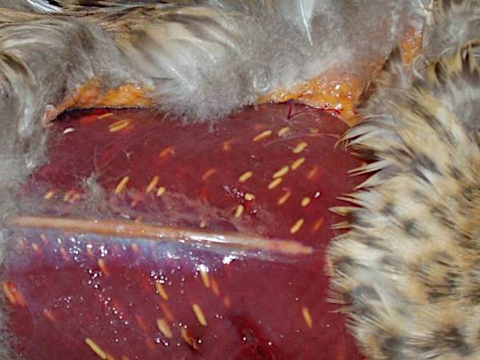
Infections are marked by the production of multiple sarcoquistes, which are sacs that contain parasites, ranging in size from micrometers (µm) to several inches, and are located in the muscles or in the nervous tissue of several animal species.
The genus includes more than 100 species show differences in their level of pathogenicity, which depend on the host specificity, as well as the structure and the place of the sarcoquistes. Have a typical life cycle of coccidios, which develops in three stages: merogonia (asexual multiplication-division multiple), gametogonia (sexual phase) and sporogonic (which is to infect new hosts).
In general, the disease is asymptomatic, given that its development is chronic and, in its initial phase, the changes do not impact directly on the muscle fibers, but the intercellular space. The occurrence of lameness and stiffness is observed only in severe cases.
Sarcocystis horvathi acts as a protozoan parasite-type intracellular. The definitive hosts of this parasite are carnivores or predators, such as dogs, cats, foxes, coyotes, and even humans. Birds, specifically chickens and ducks, working as a host intermediates to become infected by eating oocysts of S. horvathi or esporoquistes excreted through the feces of carnivores or predators.
The sarcosporidios are more frequently found in certain species duckssuch as:
In contrast, we observe a different behavior in ducks that feed on aquatic vegetationfor example:
This phenomenon is related to the differences in the manner of feeding of these birds. In addition, it has been shown that the pathological changes, which involve the analysis of the characteristics of a tissue sample, they are more frequent in adult birds (over a year), suggesting a prolonged incubation period of the disease.
The ducks, in their role as hosts intermediates, are infected through the digestive system, by means of the intake of plant foods, water, or any drink that is contaminated. It is likely that transmission occurs through drinking water, ponds, or areas contaminated shallow inhabited by predators that contain oocysts ooculados. Consequently, their main hosts intermediates are, in their majority, wild ducks.
In the digestive tract of ducks, the membrane of the ooquiste features a wrap-extremely fragile at the moment of the separation of the host cell, or during the passage through the intestine, breaking it with ease. This process allows it to be digested and that the sporozoites are released, entering the epithelial cells of the intestine.
From this point, after multiple divisions, are transformed and penetrate the epithelial layers in search of capillary blood vessels that facilitate their spread throughout the circulatory system. The merozoites become metrocitos in the muscles, which are then converted into cistozoitos, which in turn evolve to sarcoquistes.
Birds affected rarely show clinical signs, being only the ducks in an advanced state of invasion which may experience difficulties in the flight, which is related to the damage in the pectoral muscles caused by a strong attack of the parasite.
This parasite impairs flight ability, making it easier for predators to capture the ducks, which in turn allows the parasite’s life cycle to continue. In the intestines of the definitive hosts, the parasite reproduces exclusively through sexual reproduction; therefore, the intestinal walls do not suffer significant damage and show no obvious clinical signs. The only symptom that might be observed is brief diarrhea.
The life cycle of the parasite includes a host intermediate for the asexual stage and a definitive host for its sexual phase, commonly a herbivore and a carnivore in the dynamics of predator-prey. Initially, we carried out a series of stages of asexual reproduction, which end in the creation of sarcoquistes.
The ingestion of infected tissue cysts by the definitive host causes the sexual reproduction of the parasite in your digestive tract, followed by the excretion of infectious forms through the feces. The cycle is completed when a guest intermediate is infected by the mechanism fecal-oral.
In laboratory environments, it has been proven the possibility of infection in several species of birds, decorative, such as canaries, fins, australian, parakeets, pigeons, and domestic fowl, Guinea.
These birds were inoculated orally with esporoquistes of Sarcocystis obtained from opossums infected experimentally. All the birds, except for the Galliformes, showed susceptibility to the merogonia, which is a phase of the life cycle of the parasite. The pigeons were susceptible to the merogonia early, although not to the stages that affect the muscles. In contrast, the Passeriformes and Psittaciformes were fully susceptible, and the parasite became cysts in muscle in your body.
In relative terms, the invasion of these parasites in birds that bite (Galliformes) are rare, with 6 cases in chickens and 2 in wild turkeys in the southern United States.
Gracey also mentions birds among the affected species. The cysts are found within or between muscle fibers, taking on the distinctive shape of a cigar. The incidence is higher in older birds, those raised in more intensive conditions, and those that consume waste more frequently as food.
The sarcocistos that are damaged or altered may induce foci of myositis eosinofílicas that come together to form lesions of several centimeters in diameter. In the case of the pheasant, in the Czech Republic, records the presence of Sarcocystis horwathi.
Sarcocystis with forms of cigarette, in a mammal, the host end.
Literature review:
Sarcocystis (2008). En: Mehlhorn H. (eds.) Enciclopedia de parasitología Springer, Berlín.
MERCK & CO. (1995). Manual Merck de Veterinaria. Rahway, N. J., EE. UU.
BUXADÉ, P. (1987). The laying hen. Ed. Mundiprensa. Madrid.
DORN, P. (1987). Manual of avian pathology. Ed. Acribia. Zaragoza.
Betlejewska K., Kalisińska E., Kornyushin V., Salamatin R. 2002. Eucoleus contortus (Creplin, 1839): Nematodo en el ánade real (Anas platyrhynchos Linnaeus, 1758) del noroeste de Polonia. Revista electrónica de universidades agrícolas polacas.
HOFSTAD, M. S. (1984). Diseases of Poultry. Iowa State University Press, Ames, Iowa.
ZARZUELO, E. (1982). Vade mecum of the pathology, infectious poultry. Ed. Aedos, Barcelona.
CASTELLÓ, F. and CASTELLÓ, J. A. (1960). The New Art of Raising Chickens. Aedos, Barcelona.
Estudio comparativo de la fauna de cestodos y nematodos del tracto gastrointestinal de los patos silvestres (Anas platyrhynchos L. 1758) de tres ecosistemas polacos diferentes. Acta Sci. Polonorum Zootechnica. Prakas P., Liaugaudaitė S., Kutkienė L., Sruoga A., Švažas S. 2015
OROZCO, F. (1989). Breeds of chickens Spanish. Ed. Mundiprensa. Madrid.
LACADENA, J. R. (1998). Genetics. Ed. AGESA
Williams, N. A., Calverley B. K., Mart J. L. 1977. Parasites blood of wild ducks and ducks painted to the centre of Alberta and the Mackenzie Delta, Northwest Territories.
PUERTAS, M.J. (1992). Genetics: Fundamentals and Perspectives. McGraw-Hill Interamericana.
Sarcosporidiosis en patos. Parasitología Sokół R., Ras-Noryńska M.A., Gesek M.A., Murawska D., Hanzal V., Janiszewski P.E. 2016.
SANCHEZ-MONGE, E. (1969), Genetics. Espasa-Calpe S.A.
OROZCO, F. and ROBLA, F. (1986). Genetic aspects of the León rooster. 24th Symposium of the WPSA (Spanish Section): 199–212.
Hoppe, D. 1976. Prevalence of Sarcocystis detectable macroscopically in ducks of North Dakota. J. Diseases of wildlife.
Kavetska K. M. 2008. Nematofauna of duck of the genus Melanitta (Mergini, Anseriformes) from the southern Baltic sea. Wiad Parazyt.
HILL, J. L. (1973). Genetics, general and applied. Ed. UTEHA.
CASTELLÓ, J. A., LLEONART, R., FIELD, J. L., OROZCO, F. (1989). Biology of the chicken. Real Escuela de Avicultura.
Nowak M., Kavetska K., Królaczyk K., Stapf A., Korna S., Wajdzik M., Basiaga M. 2012. Estudio comparativo de la fauna de cestodos y nematodos del tracto gastrointestinal de los patos silvestres (Anas platyrhynchos L. 1758) de tres ecosistemas polacos diferentes.
LLEONART, F., ROCA, E., CALLÍS, M., GURRI, A., PONTES, M. (1991). Poultry Hygiene and Pathology. Royal School of Poultry Science.
STURKIE, P.D. (1968). Fisiología aviar, ed. Acribia. Zaragoza.
LOHMANN ANIMAL HEALTH (2012)
Sokól R., Ras-Noryńska M. A., Gesek M. A., Murawska D., Hanzal V., Janiszewski P. E. 2016. The parasites of the mallard (Anas platyrhynchos) as an indicator of health status and the quality of the environment.

You may be interested in these other sections