The chicken respiratory system is one of the most efficient in the animal kingdom. Unlike mammals, chickens use a unique airflow system that keeps fresh air moving continuously through the lungs, improving oxygen exchange and overall efficiency.
This adaptation is essential in birds and is closely linked to their evolutionary origins, tracing back to ancient reptiles and dinosaurs.
In this article, you will learn how a chicken breathes step by step the main parts of its respiratory system, and why it works in such an efficient and unique way.
Chickens don't breathe the same way humans do: Their respiratory system is much more efficient and sophisticated: Inside the anatomy of the chicken, the respiratory system plays an essential role; it is designed for flight (even though they do not fly often) and for a high metabolic rate, as we will see later.
Unidirectional airflow: In birds, air flows through the lungs in a single direction (unidirectional), which means that these organs are often supplied with fresh air, whether they are inhaling or exhaling.
The lung does not expand: In humans, the lungs expand and contract. In chickens, the lungs are rigid and do not change in size; instead, it is the air sacs that expand and contract to circulate air.
Air bags (similar to bellows): Chickens have nine air sacs distributed throughout their bodies that act like bellows, storing and moving air, unlike humans, who use lungs to inhale and exhale.
The process of breathing: Inhalation: Air enters through the nostrils, passes through the trachea, and first reaches the posterior air sacs, not the lungs. Exhalation: During exhalation, air moves from the air sacs (alveoli) to the lungs to facilitate gas exchange.
It is important to note that, among vertebrates, birds are the group that has been most successful in conquering the skies. Their respiratory adaptations are undoubtedly the key to this success.
The chicken respiratory system is a specialized set of organs designed to maximize oxygen intake and efficiency. Unlike mammals, chickens rely not only on their lungs but also on a system of air sacs that allows air to flow continuously through the body.
This system is made up of several structures that work together to ensure a constant supply of fresh air and an efficient exchange of gases.
Each of these components plays a key role in the breathing process.
Chickens do not breathe in the same way as humans. Their respiratory system is more efficient and works through a continuous airflow process that allows fresh air to pass through the lungs during both inhalation and exhalation.
Step-by-step process:
This continuous airflow system allows the lungs to receive fresh air at all times, making the chicken respiratory system far more efficient than that of mammals.
Air sacs are one of the most important features of the chicken respiratory system. They act as reservoirs that store and move air throughout the body, allowing a continuous and efficient airflow.
Functions of air sacs:
Chickens typically have nine air sacs distributed throughout their body, and some of them even extend into certain bones, further enhancing the efficiency of the respiratory system.
The efficiency of the chicken respiratory system lies in its one-way airflow. Unlike mammals, where air moves in and out of the lungs, chickens maintain a continuous flow of fresh air, which greatly improves oxygen absorption.
This means that chickens can extract more oxygen with less effort, making their respiratory system highly efficient. Although this adaptation is essential for flying birds, it also provides important benefits for domestic chickens.
This system is especially important for birds that fly, but it also benefits domestic chickens.
The respiratory system of modern birds, including chickens, has its origins in ancient theropod dinosaurs. Fossil evidence suggests that these dinosaurs already had structures similar to air sacs, allowing them to breathe efficiently.
In addition to this respiratory adaptation, birds still retain other evolutionary traits, such as the scales on their legs, which clearly show their connection to reptiles.
Another evolutionary trait that birds share with reptiles is the presence of scales on their legs, a visible reminder of their ancient origins.
A bit of history. The dinosaur ancestors of today's birds likely breathed in a similar way to their modern descendants. This has been revealed by a study of the fossil remains of a "Majungatholus atopus," a theropod dinosaur related to Tyrannosaurus rex.
The findings of the study, conducted by researchers at the Ohio College of Osteopathic Medicine (United States), have been published in the journal *Nature*.
Researchers have discovered evidence that in the dinosaurs' theropods, a group that belongs to the Tyrannosaurus, Velociraptor, or the Carnotaurus, they pumped air into bags hollow in the interior of the skeleton, as the majority of birds.
Scientists say that a new fossil of *Majungatholus atopus*, an ancient theropod that could grow several meters long and is distantly related to Tyrannosaurus rex, shows that these dinosaurs had everything necessary for this type of respiration.
Scientists explain that the vertebrae of the creature, between 70 and 65 million years ago, show adaptations for respiration similar to those of the crane and white-tailed deer, indicating that the system did before the birds today.
Scientists also explain that birds maintain a highly active lifestyle by using a series of additional air sacs that provide their lungs with a constant supply of oxygen-rich air, rather than relying on a single inhalation and exhalation cycle as mammals do.
According to the researchers, because these bags are located in the empty spaces between the bones, they have found evidence of their existence in dinosaur theropods. However, until now, scientists were not sure that the system was sophisticated enough to work in the same way as in modern birds.
The respiratory system differs considerably from that of mammals, as it has no diaphragm, and breathing is active (with the expenditure of energy) and, therefore, a need for ventilation, powerful and fast. This act, locomotion, demands a great muscular effort, resulting in a high consumption of oxygen.
Well, we're going to analyze these features, following the anatomical structure of the respiratory hen.
A healthy respiratory system is essential for a chicken’s overall well-being.. Poor ventilation, dust, or high humidity levels can negatively affect a chicken’s respiratory system and lead to serious health problems.
Maintaining good environmental conditions is essential to prevent respiratory issues and keep chickens healthy and productive.
Practical recommendations:
The air you need our chicken's nasal cavity is accessed through the nostrils located at the base of the beak. Let's take a look at the horns and breasts.
The nasal cavities are separated by a thin cartilaginous nasal septum, which may be incomplete rostrally (permeable nose in web-footed birds). There are three nasal conchae (rostral, middle, and caudal), and their development varies by species. The ethmoid labyrinth is not mentioned because the hen’s sense of smell is poorly developed.
Of the paranasal sinuses, only the infraorbital sinus is present, which corresponds to the maxillary sinus in mammals. This sinus is located ventrally and caudally to the eye and communicates with the caudal nasal concha and the nasal cavity itself. In psittacines, the left and right infraorbital sinuses are connected and have diverticula that fill large areas of the skull with air. Upper respiratory tract infections can affect these sinuses, where pus accumulates and is difficult to treat.
The nasal turbinates are highly vascularized, as they are composed of an extensive network of blood vessels (rete mirabile) that play a role in thermoregulation and homeostasis (which, to put it simply, refers to the balance within the internal environment; the hen’s body mounts adaptive responses to maintain health), just as water is essential for our birds. Rehydration of the hen can be promoted during very hot periods or in very hot areas through aerial misting.
Already the air in the nasal cavity runs through the choana in the direction of the oropharynx, reaching the glottis.
The nostrils They open in the dorsal groove of the beak, either in the horny part or in the cere (a transitional skin tissue rich in nerve endings). They are located at the base of the beak and are covered by a protruding horny flap called the operculum.
There is an intermediate septum, and it communicates with the oropharynx via a choana, as in mammals. It has rostral, middle, and caudal nasal turbinates. The nasolacrimal duct is wide and opens into the ventral nasal cavity and the middle nasal turbinate
From the choanal (that are opened directly in the hard palate), the air passes into the trachea, through the larynx, integrated only by the cartilages arytenoides and the cricoid (no epiglottis).
The glottis is situated on a small hill known as the laryngeal prominence. Nor are there in birds, vocal folds, and the muscles of the larynx; they are very rudimentary. This is because the larynx, unlike in mammals, is not involved in the emission of sounds (phonation).
Rises in the oropharynx, forming the prominent larynx. It consists of the cricoid cartilage and two arytenoid cartilages, which form the glottis. Like other systems in the body, as is also the case in the the hen's digestive system, where each organ has a specific function.
During swallowing, the two arytenoid cartilages come together and close off the entrance to the larynx, since there is no epiglottis or vocal folds. Sounds are produced in the syrinx.
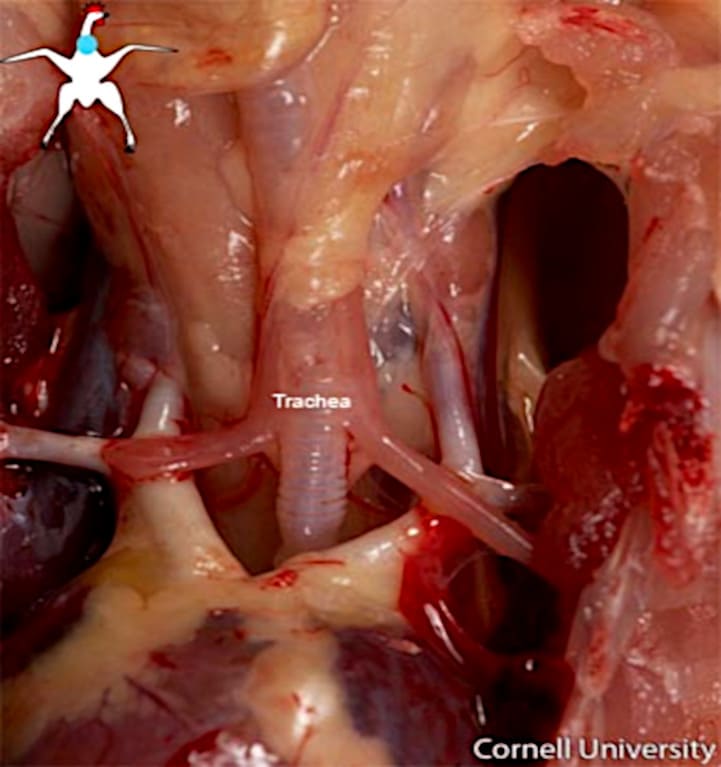
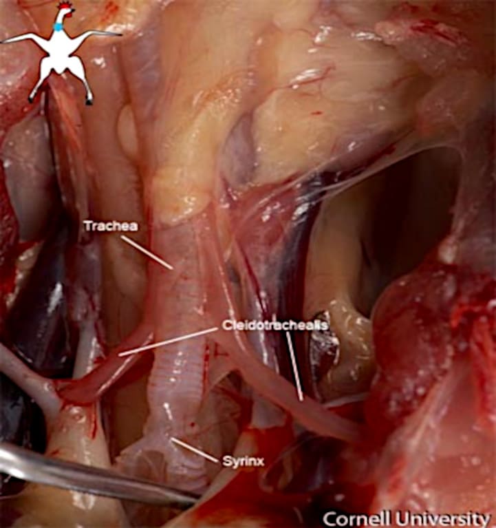
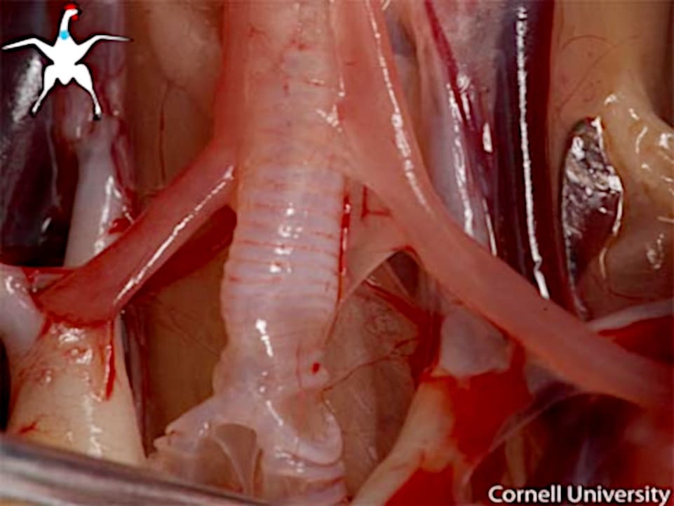
Source; Photos of Cornell University
The trachea of birds has a number of features that must be known; it is long and is composed of rings of rigidity and completeness, presenting very different ways.
The hens have a trachea, a rigid structure, and a greater length and diameter than the mammals of similar sizes. The birds have increased the diameter of the trachea, or windpipe, to compensate for the length; this way, the resistance of the trachea to the gas flow is similar to that of mammals.
In the world of birds, their anatomy features a variety of tracheal structures, including complex and contoured forms. For example, we can cite the double tracheas of penguins, the emu’s trachea in running birds with a diverticulum that opens into the cervical trachea, the tracheal bulb found in male ducks, and spiral or loop-shaped tracheas, such as those of swans, spoonbills, or cranes.
The trachea consists of 100 to 130 cartilage rings that tend to ossify. Such rings are also present in the main bronchi.
In species of long neck (swans, cranes), on the left side of the sternum, which features a notch at the end, the trachea describes gyri (hypertrophy tracheal). Such hypertrophy seems to be related to an increase of the power of the voice.
The bifurcation of the trachea is modified to form the true vocal organ of birds: the syrinx, or caudal larynx, which may be absent in certain species (vultures, ostriches, and some storks). It is composed of complete cartilaginous rings that can be felt on the right side of the trachea.
The trachea branches into two main bronchi located dorsal to the base of the heart, which then pass through the ventral surface of the lungs.
Birds have a higher tidal volume (the amount of air displaced during a normal inhalation and exhalation) and a slower, deeper respiratory rate than mammals of the same size. For example, a 300-gram pigeon breathes 26 to 29 times per minute, whereas a rat of the same weight breathes up to 85 times per minute.
It is the organ vocal of the birds and is well developed in some species. It is characterized because it is a narrowing, located between the trachea and the bronchi, facilitating, regardless of their sounds or songs, that is a usual site of obstruction by foreign bodies, like a seed of oats or barley, producing a characteristic sound in the chicken when you try to delete it; look at it.
Formed by the end portion of the trachea, or windpipe, and the initial segment of the main bronchi. Cartilage tracheal area corresponding to the syrinx, are strong, while in the bronchial practically are absent in this region.
The lateral and medial walls of the bronchi are membranous and produce sound when they vibrate. Male ducks and swans have a bony bulla, which acts as a resonating structure, on the left side of the syrinx.
The syrinx is equipped with powerful muscles that tense a vibrating membrane. The quality and complexity of song depend directly on these muscles. For example, hawks, which produce only a few calls, have only two pairs of muscles, while passerines (such as canaries) have as many as seven or nine pairs.
They are small, without lobes, bright red in color, soft. Located beneath the thoracic vertebrae and ribs, which create indentations or depressions on the dorsal surface of the lungs. From a morphological standpoint, the lungs of birds account for less than 15% of a hen’s respiratory system and are connected to the air sacs. However, their role in gas exchange is minimal, contributing only 5% of the total.
Birds' lungs are more rigid and do not undergo the changes in volume seen in mammals. Even so, despite the small volume of their respiratory system, they have a large functional capacity due to a number of unique characteristics that we will discuss below.
The lungs do not cover the lateral surface of the heart, as they do in mammals; there is no pleural cavity, and lung expansion is limited. The primary bronchi through the lungs in all their length (mesobronchi), ending in the air sacs of the abdomen.
In the hen's lung, each primary bronchus divides into four secondary bronchi, which in turn form tertiary bronchi known as parabronchi. This is the area where gas exchange takes place due to a complex network of air capillaries, which are closely connected to blood capillaries.
These capillaries of air, where the gas exchange occurs, are of smaller size (3-10 µm) than the alveoli of mammals (>35 µm) and form a three-dimensional network of airways, in that the air flows in a single direction.
The number of tertiary bronchioles varies by species, with the number increasing as the bird becomes more specialized for flight. In birds, the bronchioles consist of a group of hundreds of parallel tubules that make up what is known as the paleopulmon.
In some species, there is also a less prominent pair of bronchioles with irregular branching, known as the neopulmon. Some birds, such as the kiwi, the emu, and the penguin, lack this structure, while it is minimal in pigeons (10–12%) and more prevalent in passerines and psittacines (20–25%).
The air passing through the paleolung always moves in the same direction, whether during inhalation or exhalation, but in the neolung, the direction depends on the respiratory phase, and the movement of air is bidirectional.
Source: Photos of Cornell University
The main bronchi:
They penetrate the lungs through the ventral side, pass through the lungs, and continue at the caudal edge, each with an abdominal air sac.
In the chicken, each main bronchus emits 40 to 50 bronchi side-classified as:
There are four medioventral bronchi originating from the main bronchus after entering the lung.
The first one gives off a branch to communicate with the cervical air sac; the third one communicates with the clavicular and cranial thoracic air sac.
Of the bronchi lateroventrales, one of them is connected to the air sac's thoracic flow.
The bronchi side generated from 400 to 500 parábromquios, on whose walls he performs gas exchange.
The set of these parabronchios generates the portion of the functional lung called paleopulmón.
The extensions of the light to the bronchial generate the capillary air, which creates a network of handles interconnected.
These capillaries are intertwined with blood capillaries, leading to the main part of the wall parabrónquica, a site and structure where the gas exchange occurs.
Air capillaries are the mammalian equivalents of pulmonary alveoli.
Air sacs of a Chicken (as contrasts)
1 – Pulmo dexter; 1 - Incisurae costales; 2 – Sacci cervical; 2 - Ductus intertransversarius;2 – Diverticulum supramedullare; 3 – Saccus Clavicularis; 3 – Diverticulum axillare; 3 – Diverticulum subcordale; 3 – Diverticulum humerale; 4 – Saccus thoracalis cranialis; 5 – Sacci thoracales cudales; 6 – Sacci abdominal; 6 – Diverticula gastric; 6 - Diverticula acetabularia; 6 - Diverticulun iliolumbale; 7 – Humerus; 8 – Foramen pneumaticum; 9 – Trachea.
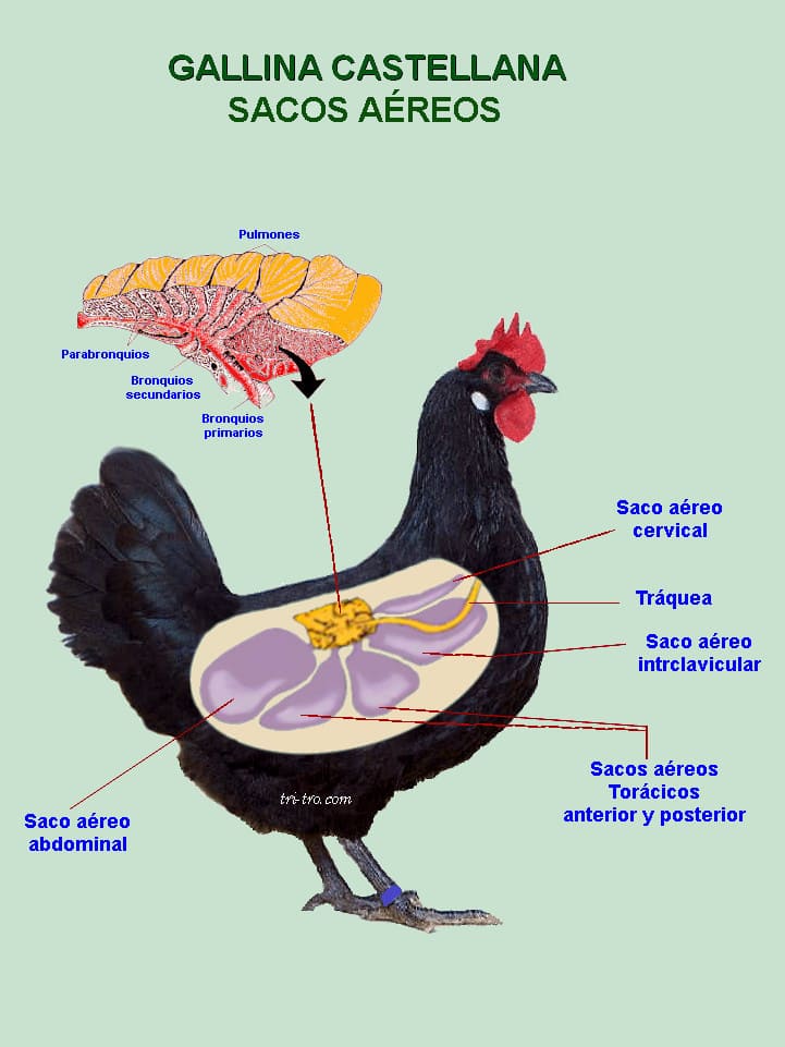
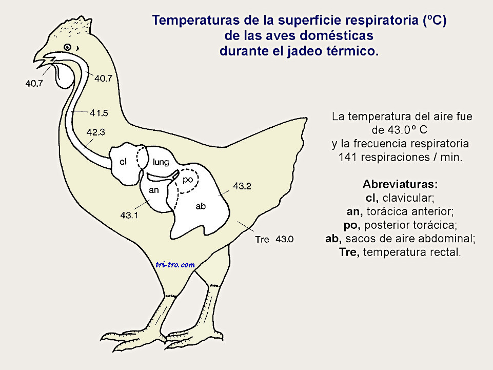
These bags play a vital role in the breathing of the hen, as they come to represent 80% of the volume of the bird's respiration. The air sacs act as bellows or a pump system that streamlines the flow of air over the surface effectively, unlike the lung, which is not expandable.
Are dilations of very thin bronchial systems extended beyond the lungs in relation to the viscera, thorax, and abdomen? To be formed by a thin-walled avascular tissue (which contains no blood vessels, so it has no blood supply of its own), this makes them difficult to access for the drugs when injected intramuscularly. The infections are frequent and difficult to treat.
Some diverticula of these way into several of the bones, giving them the feature of bone tires, such as the femur, humerus, sternum, cervical vertebrae, and the bones of the wings, but not in all species. A wound or injury that may occur in these bones or tires can lead to infection in both the lungs and air sacs.
The primary function of air sacs is to reduce body weight to facilitate flight and swimming, but they also prevent the body from overheating during these activities. For this reason, any problem can seriously affect the animal’s health.
The hen has eight air sacs; the cervical and clavicular air sacs, which connect to the ventral bronchus, are unpaired, while the cranial thoracic, caudal thoracic, and abdominal air sacs, which connect to the primary bronchus, are paired. In exotic birds, the cervical air sac may be double.
The air sacs are also very present in the vocalization, the courtship, and the thermoregulation of the testes during spermatogenesis (the process of the formation of the male sex cells, from spermatogonia to spermatozoa).
Cervical sac: Presents diverticula extending throughout the cervical and thoracic vertebrae.
Clavicular sac: larger in size, located at the entrance to the chest, extending to the sternum.
Cranial thoracic sacs: located ventrally to the lungs, between the ribs, sternales, the heart, and the liver.
Bags thoracic flows: located in the flow rates between the body wall and the abdominal bags.
Abdominal sacs: are the largest, located caudodorsales to the abdominal cavity in contact with the intestines, gizzard, genital organs and kidneys. Their diverticula penetrate to the sacrum and the acetabulum.
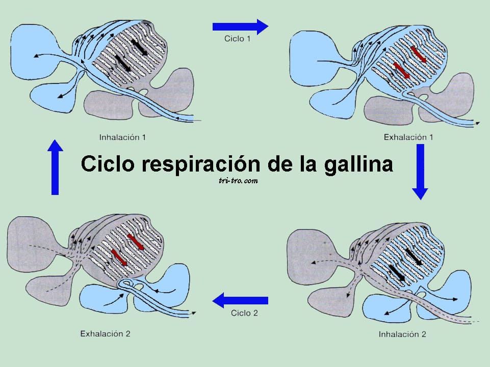
The respiratory cycle in birds is very complex, and then we will make only a simplification of the same, trying to explain the phenomenon of inspiration and expiration.
Air capillaries are the mammalian equivalents of pulmonary alveoli.
We first go to the definition that comprises this process. Inhalation (the act of drawing air or another gaseous substance into the lungs) and exhalation (the expulsion of air from the lungs; it is, therefore, the opposite of inhalation).
Inspiration: The intercostal, serratus, and scalene muscles contract; the ribs move forward and the sternum downward.
Air passes through the trachea, then into the primary bronchi, and then into the caudal air sacs (caudal thoracic and abdominal). At the same time, air from the previous respiratory cycle enters the lungs, then into the cranial air sacs (cervical, interclavicular, and cranial thoracic).
Expiration: In this second phase of the respiratory cycle, the abdominal muscles contract, the air sacs are compressed, the air from the caudal sacs is transferred to the lungs for gas exchange, and the air from the cranial sacs is sent to the primary bronchi and trachea.
As a result, pulmonary ventilation occurs more freely and thoroughly than in mammals. Since birds lack a muscular diaphragm, their inspiratory and expiratory movements depend on the muscles of the thoracic wall; this active muscular involvement drives airflow through the paleopulmon, always moving from caudal to cranial.
When a chicken inhales during the first cycle, it does not exhale completely as we do; it must exhale during the second cycle. In this way, 50% of the air inhaled during the first cycle passes through the lungs, via the primary bronchus, to the caudal air sacs.
The other 50% goes to the lungs, where gas exchange takes place, following the air that enters the paleopulmonary system from the posterior air sacs during exhalation in the first cycle. In addition, it is important to monitor the hens’ overall health, as they may suffer from other health issues, as explained in our article on how to treat wounds in hens.
During the inhalation phase of the hen’s second respiratory cycle, this air enters the cranial air sacs, from which it is eventually expelled during the exhalation phase of the second cycle.
This makes the air of the lungs of the chicken will renew both in inspiration and in expiration of each cycle. Birds lack a diaphragm, so that your breathing is based on the movements of the intercostal muscles, external and internal, and the abdominal muscles.
The lungs are rigid and do not act in the process of mechanical ventilation, so you do not have to expand or contract as mammals do, having little interstitial tissue to have greater resistance. This way, your area of gas exchange in the hen is 20% greater than in mammals.
Respiratory infections in chickens are common these highly contagious diseases are generally caused by viruses (such as bronchitis, laryngotracheitis, or Newcastle disease) or bacteria (such as coryza or mycoplasma). Symptoms include coughing, sneezing, hoarseness, nasal and ocular discharge, and shortness of breath. Vaccination and biosecurity are essential to prevent high mortality and declines in egg production. Other factors can also exacerbate infections. open wounds.
Most common respiratory diseases:
Common symptoms we have observed:
Control and prevention:
To take into account: It is advisable to visit your veterinarian if any suspicious symptoms appear in order to obtain an accurate diagnosis, since many conditions have similar symptoms.
For all of the above reasons:
The hen's respiratory system is a highly efficient mechanism that allows these birds to obtain more oxygen than mammals. Hens are able to maintain optimal performance levels, even under challenging conditions, thanks to their air sacs and their dual air circulation system.
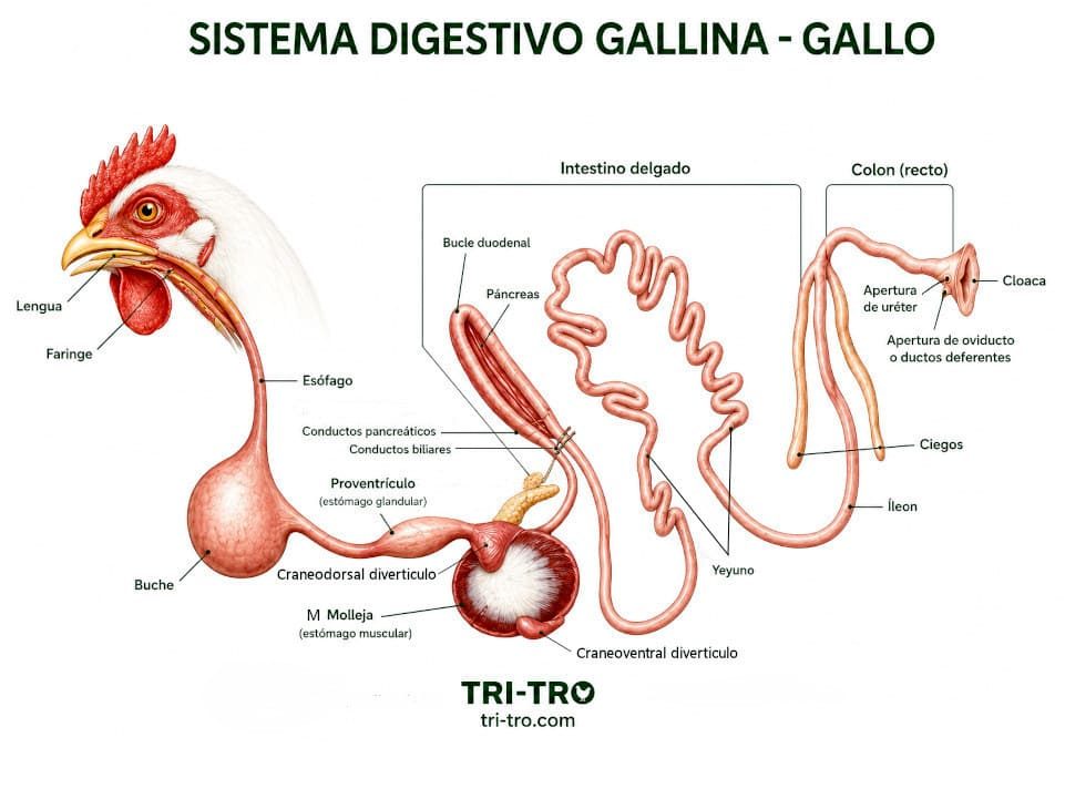
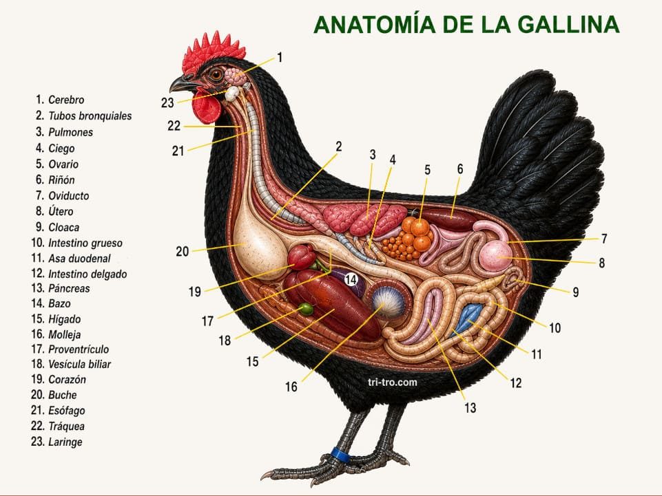
To better understand how a bird's body works, you can also look into other systems, such as the digestive system, or the complete anatomy of a hen.
To provide better care for chickens, prevent respiratory diseases, and ensure their well-being, it is essential to understand how they breathe. In addition to respiratory diseases, chickens can also suffer injuries, as you can see in our article on injuries in chickens.
The respiratory system of the chicken is a remarkable example of evolutionary adaptation. Its unique airflow mechanism, supported by air sacs, allows for efficient oxygen exchange and plays a crucial role in the health and vitality of the bird.
Understanding how it works helps poultry keepers provide better care and prevent respiratory issues.
Chickens breathe using a two-cycle airflow system that allows air to pass through the lungs twice, increasing oxygen efficiency.
Air sacs are structures that store and move air through the body, ensuring continuous airflow and helping regulate temperature.
They function like bellows that store air, ensuring that the lungs receive fresh oxygen both when exhaling and inhaling.
Because it maintains a constant flow of fresh air through the lungs, improving gas exchange compared to mammals.
Bibliography:
Dyce, K. M., Sack, W. O., and Wensing, C. J. G., 1991. Veterinary Anatomy. Ed. Pan-American.
Ede, D. A., 1965. Anatomy of birds. Ed. Acribia.
Grasse, P. P., 1980. Zoology. Vol. 4: Vertebrates. Reproduction, biology, evolution, and systematics. Ed. Toray-Masson.
Grifols, J. and Molina, R. 1994. Manual of clinical exotic birds. Ed. Grass-Iatros.
Natural History, 1984. Vol. 1. Ed. Ocean-Institute Gallach.
King, G. M. and D. Custance. Colour atlas of vertebrate anatomy: The Pigeon. Pp.: 5.1— 5.17. Ed. Blackwell Scientific Publications.
Krahmer, R., and Schröeder, L. 1979. Anatomy of the domestic animals. Ed. Acribia.
McClelland, J., 1992. Atlas in color of the anatomy of birds. Ed. inter-American.
Nickel, R., 1977: The Anatomy of the Domestic Birds. Ed. Paul Parey.
Sandoval, J., 1976. Veterinary Anatomy. Ed. Modern Printing Press (Córdoba).
Schwarze E. 1980. Compendium of Veterinary Anatomy. Volume V: Anatomy of birds. Ed. Acribia.
Shively, M. J., 1993. Veterinary anatomy, basic, comparative, and clinical. Ed. Manual Moderno.
Sturkie PD, 1967. Physiology of Avian. Ed. Acribia.
Dr. Francisco Gil Cano.
Professor H. Aguinaga.
Barsotti G, Briganti A, Spratte JR, Ceccherelli R, Breghi, G. (2010). Bilateral mydriasis in common buzzards (Buteobuteo) and little owls (Athene noctua) induced by concurrent topical administration of rocuronium bromide. Veterinary Ophthalmology, 13 Suppl: 35-40.
Barsotti G, Briganti A, Spratte JR, Ceccherelli R, Breghi, G. (2012). Safety and Efficacy of Bilateral Topical Application of Rocuronium Bromide for Mydriasis in European Kestrels (Falco tinnunculus). Journal of Avian Medicine and Surgery 26: 1-5. Calder, WA. (1968).
Respiratory and Heart Rates of Birds at Rest. The Condor, 70: 358-65. Clippinger TL. (1997).
Diseases of the Lower Respiratory Tract of Companion Birds. Seminars in Avian and Exotic Pet Medicine, 6: pp. 201-8. Dubé C, Dubois, R, Struthers, J. (2011).
Intravenous and Intraosseous Fluid Therapy in Critically Ill Birds of Prey. Journal of Exotic Pet Medicine, 20: 21-6. Daan S, Masman D, Groenewold, A. (1990).
Avian basal metabolic rates: their association with body composition and energy expenditure in nature. American Journal of Physiology – Regulatory, Integrative and Comparative Physiology, 259: R333-40.
Francis N, Ruiz Trojan JD, Agüera EI. (2003).
Lead and lead toxicity in domestic and free living birds. Avian Pathology 32, 3-13. Dorrestein GM, Van Miert ASJ. (1988).
Pharmacotherapeutic aspects of medication of birds. Journal of Veterinary Pharmacology and Therapeutics, 11, 33-44.

You may be interested in these other sections