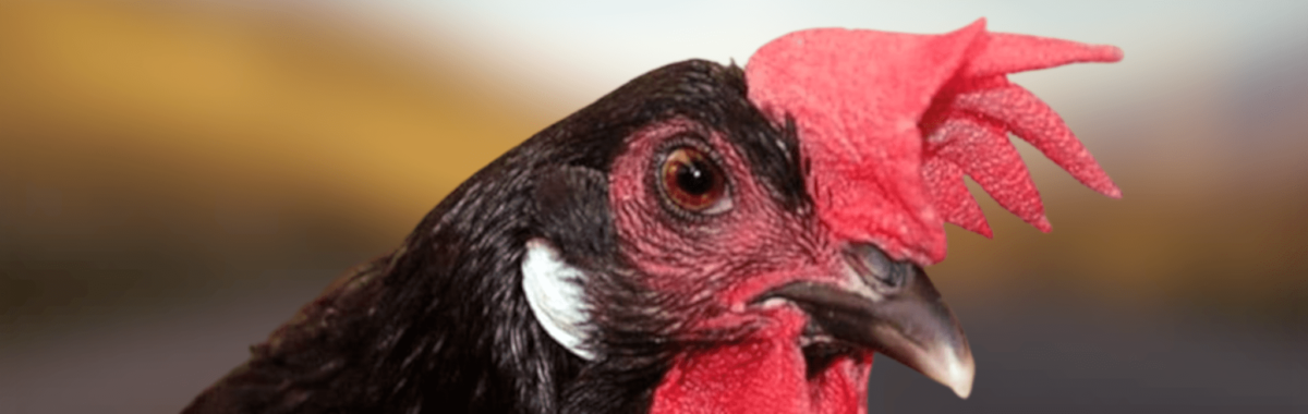
The penis of the chickens are suicidal prior to hatching

El estudio realizado

A genetic study reveals el mecanismo que hace que muy pocas aves tengan pene. Estudio realizado en la Universidad de Florida desvela el mecanismo genético que indica que muy pocas aves tengan falo. Ver vídeo al final del post para su comprensión. 

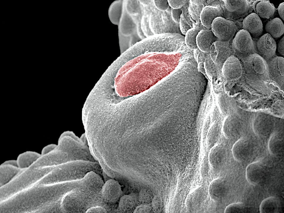
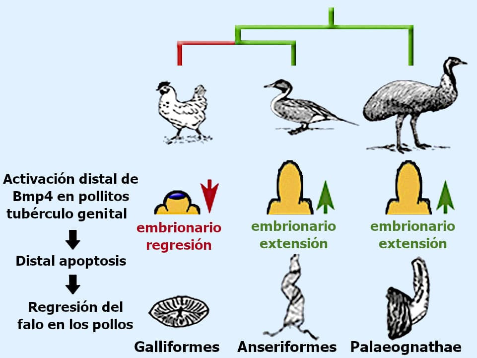
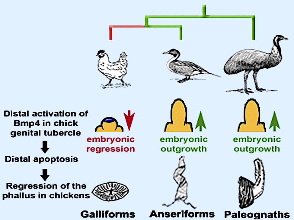
En el transcurso del crecimiento del embrión, los pollos empiezan a desarrollar un tubérculo genital, que es el predecesor del falo, ocurriendo que en un cierto momento se activa un proceso de muerte celular llamado apoptosis (es una vía de destrucción o muerte celular programada o provocada por el mismo organismo, con el fin de controlar su desarrollo y crecimiento) y el órgano empieza a encogerse hasta desaparecer antes de que el ave salga del cascarón.

Highlights

  • During the evolution of birds, the phallus was reduced or lost in most of the lineages.
  • Chickens are a phallus reduced, but retained the mechanisms of primitive growth.
  • Un dominio Bmp4 derivado provoca apoptosis y regresión del falo embrionario de los pollitos.
  • The birds and basal-ducks lack of manifestation distal Bmp4, being able to develop phalli elongated.

Background

Uno de los hechos más desconcertantes de la evolución es la reducción y pérdida del falo en las aves.

All birds reproduce by internal fertilization, but only 3% of them have retained a phallus with a capacity of penetration.

Several hypotheses have been proposed for the evolutionary mechanisms that led to the reduction of the phallus; however, nothing is known about the developmental mechanisms that underlie it.

Pocos caracteres morfológicos en el reino animal rivalizan con la diversidad, complejidad y evolución de los genitales externos masculinos. Entre los animales con fertilización interna, la morfología genital evoluciona rápidamente; el tamaño, la forma y los detalles anatómicos de los genitales externos pueden mostrar una variación dramática.

Ejemplos más llamativos de la diversificación evolutiva, tanto de la forma como de la función genital, se encuentran en las aves. De hecho, los genitales externos están reducidos o ausentes en casi 10.000 especies de aves. Estos cambios evolutivos han dado lugar a morfologías del falo que pueden clasificarse operativamente en tres categorías generales:

  1. A phallus flashing that is capable of inserting itself during copulation (e.g., ducks).
  2. A phallus is not intermittent that is reduced, but not lost (for example, chickens).
  3. Ausencia del falo, que implica una pérdida completa de una protuberancia fálica (por ejemplo, neoaves, el grupo más grande de aves).

Aunque la mayoría de los galliformes sufrieron una reducción extrema de los genitales externos, los miembros de su clan hermano, los anseriformes, conservaron un falo intermitente, y en algunas especies estos órganos en espiral alargados pueden exceder la longitud del cuerpo.

These morphological alterations led to changes in the strategies of copulation in different lineages.

Por ejemplo, los machos que carecen de un órgano intermitente o prominente transfieren los espermatozoides mediante la aplicación de la cloaca con la de la hembra, en una maniobra conocida como el “beso cloacal”, que requiere la cooperación de la hembra; ese es el caso de nuestras gallinas.

In contrast, males with phalluses can manipulate females and even forcibly copulate with unwilling females, a behavior that is well documented in waterfowl.

Although the selective pressures to obtain morphologies of the phallus made are obvious, it is less clear how the selection might favor the reduction or loss. Several hypotheses have been proposed;  

Regression of the phallus in galliformes

Phallus regression in galliforms

  1. One of the assumptions más influyentes para la diversidad genital es la del “padlock and key”; sostiene que la rápida evolución de los genitales facilitó la especialización a través de la incompatibilidad reproductiva (es decir, solo los machos y las hembras de la misma especie pueden encajar sus genitales juntos).
  2. La segunda hipótesis: Mayr proposed that genital morphology undergoes frequent changes due to pleiotropic effects. It is unclear why neutral changes resulting from the pleiotropy (es el fenómeno por el cual un solo gen es responsable de efectos fenotípicos o caracteres distintos y no relacionados) afectarían a los genitales de manera desproporcionada, aunque los estudios del control genético del desarrollo genital han identificado una multitud de mecanismos de desarrollo (incluyendo elementos reguladores del ADN que actúan) que son compartidos por otros sistemas de órganos.
  3. A third hypothesis argues that the diversification of the external genitalia has been driven by sexual selection que actúa sobre la variación; eso afecta la competencia espermática, las características sensoriales de la morfología genital, la elección femenina y la adaptación sexualmente antagónica (es decir, los machos y las hembras evolucionan adaptaciones y contraadaptaciones para ganar control sobre la cópula y la fertilización).

The recent progress in biology on the development of the external genitalia in mammals has led to the identification of genetic pathways that control the growth and patterning of the penis. 
These discoveries, coupled with advances independent phylogeny avian reproductive ecology, and morphology, comparative, created a new opportunity to investigate the genetic basis of the development of the evolution genital in birds. 

The same mechanisms that regulate the development of the penis of mammals also operate on the external genitals of the birds, since the proliferative signals that direct the growth direct genital preserved in the embryos of chickens (a galliforme) and duck (a anseriforme) despite the striking differences in their genital adult. 

Aunque el desarrollo temprano del tubérculo genital (el precursor del falo) se conserva en pollos y patos, encontramos que los pollos tienen un dominio único de muerte celular distal. Este dominio de la muerte celular está asociado con la activación ectópica (de un gen en un tejido donde no se expresa normalmente) del gen que codifica la proteína 4 morfogenéticas ósea (Bmp4).

Evolution of the reduction of the phallus in birds

The marked divergence the differences in genital morphology between anseriformes and galliformes raise questions about the selective pressures that led to phallic reduction. The loss of intromittent organs is an evolutionary paradox: 

How can the reduction of a structure that facilitates internal fertilization have a positive effect on the reproductive fitness?

It is unlikely that the reduction of the phallus is the result for better delivery of sperm. 

Darwin proposed for the first time that the character variables, without adaptive value apparent, were not subject to natural selection, but that these traits could be stabilized in a population if confer an advantage “over other individuals of the same sex and species, in exclusive relationship with the reproduction.”

The copulation with males that lack of a phallus intromitente requires the cooperation of the female, as the presentation of the cloaca and the eversion of the vagina. Accordingly, when you select males with phallus reduced or not intromitente, females can control the paternity, which is consistent with the idea that the election of women played a role in the reduction and loss of the phallus.

Una alternativa a la hipótesis de la selección sexual es que la reducción del tamaño del falo puede haber sido un efecto secundario de los cambios de desarrollo en otros caracteres.

The molecular mechanisms of the development of the appendix have been preserved deeply during animal evolution, and gene networks that regulate the development of the external genitalia also control the formation of a wide range of other structures, including the extremities, the gut, the nervous system, the muscle, and the appendices without tegumentarios, such as feathers and scales. In addition, the specific expression of the tissue of several genes expressed in the genital tubercle.

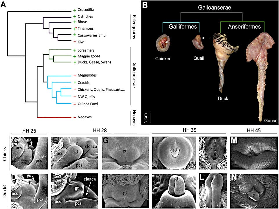
Figure 1 Comparison of phylogenetic, adult and embryology of the external genitals in birds

(A) Phylogenetic distribution of the phalli flashing in birds. + indicates present; – indicates absent or reduced in the majority of species of the classified. Most tinamous have a phallus flashing.

The colors of the branches are the following:

  • Purple, Paleognathae.
  • Green, Anseriformes.
  • Cyan, Galliformes;
  • Orange, Neoaves.
  • Black, Crocodilia (root).

(B) Anatomical preparation of external genitalia in male adults in two anseriformes (goose and duck) and two galliformes (chicken and quail). The arrows point to the reduction of the phalli in the species of galliformes.

The scale bar represents 1 cm

(C-N) Scanning electron microscopy of the development of external genital, in chicks and ducks.

Anterior is to the left in (C)-(F) and up in (G)-(N). The embryos of chickens and ducks initiate the formation of bumps genital mated ((C and D), forming a single tuber during the growth phase of early (E and H). The arrests by excessive growth and regression of the genital tubercle in the embryo of chicken (I, K y M)but genital development continues in the duck embryo (J, L y N). The numbers of stages of Hamburger-Hamilton (HH) shown above.

Abbreviations: gs, genital swelling; gt, genital tubercle; acs, swelling sewage above; pcs, swelling sewage later.

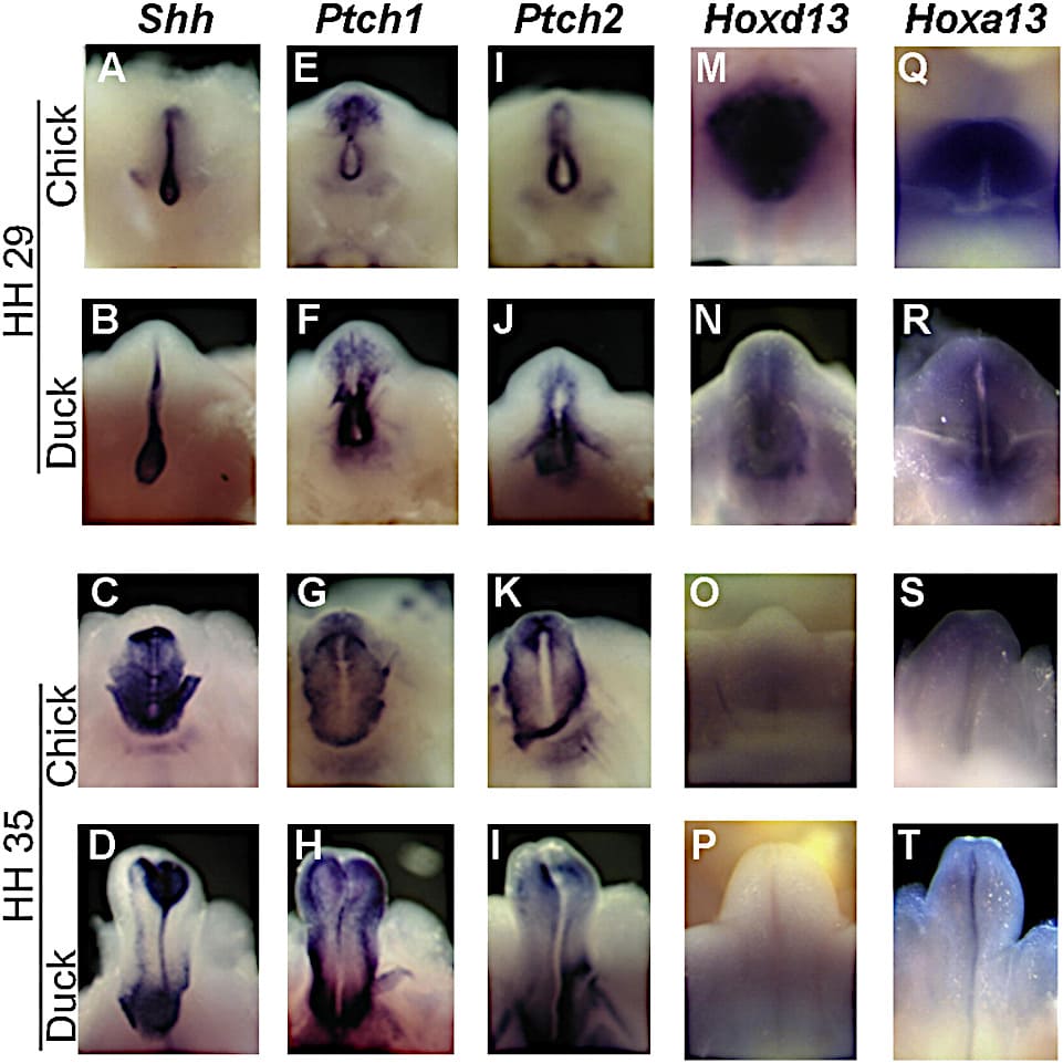
2 comparative Analysis of gene expression in the development of the external genitalia of embryos of chicken and duck

The in situ hybridizations of RNA whole of the tubers genitals of chickens and ducks in the stages 29 and 35 show patterns of expression of genes that promote the development of genital external mouse.

The tubers genitals are shown in ventral view with distal upwards.

The names of the genes are shown at the top; the species and the stage Hamburger-Hamilton (HH) are shown to the left.

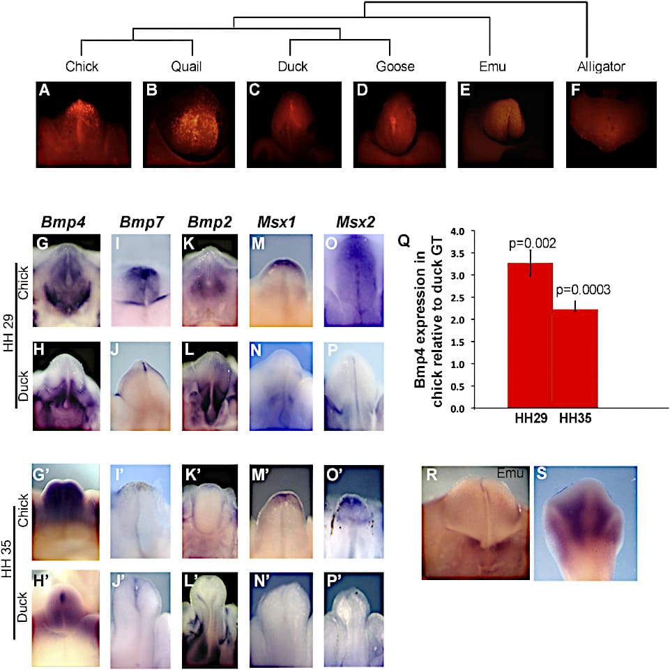
Figure 3. The apoptosis and the signaling of breast augmentation in the tubers genitals galliformes

(A-F) The red coloration of LysoTracker shows widespread apoptosis in the tubers genitals galliformes (chickens and quails), while the genitals anseriformes (ducks and geese), emus and crocodiles in stages comparable show apoptosis limited, mostly along the groove. The phylogenetic relationships are shown at the top.

(G-P) Bmp4, Bmp7, Bmp2, Msx1 and Msx2 in chickens and ducks in the stadium 29 (G-P) and 35 (G'-P').

Bmp4 (G y H), Bmp7 (I y J), Msx1 (M y N) y Msx2 (O y P) muestran expresión distal en los tubérculos genitales de los pollitos, pero no de los patos en el estadio 29 (G-J).

Los dominios de expresión distal de Bmp4 (G′ y H′), Msx1 (M′ y N′) y Msx2 (O′ y P′) persisten en los tubérculos genitales de los pollitos hasta el estadio 35. Bmp2 no muestra diferencias significativas en ninguna de las dos etapas (K, L, K′ y L′).

(Q) RT-PCR shows that the expression of Bmp4 is significantly higher in tubers genitals (GT) of the chicks in comparison with the ducks in the stadium 29 (p = 0.002) and in the stadium 35 (p = 0.0003). The error bars show the standard error of the mean.

(R) In the genital tubercle emu, the expression of Bmp4 is absent in the distal tip. The expression was restricted to the base of the tuber, and the neck sewage, as in ducks (compare H).

(S) Internal Control of the hind limb of the emu that displays the expression interdigital of Bmp4.

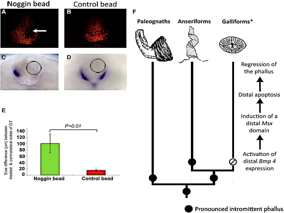
Figure 4. The signaling of Bmp cause apoptosis and growth inhibition in the genitals of the chickens.

(A-D) Las perlas se cargaron con proteína Noggin.

A y C) or PBS (B and D) and implanted on the left side (right in photo) of the tubers genitals, chick in stage 29.

(A) Noggin bead (arrow) decreases apoptosis (red blood cells are positive for LysoTracker) on the treated side of the tubers. The contralateral side shows widespread apoptosis.

(B) The account of PBS has no effect on any of the sides.

(C) Msx1 is regulated down on the side of the tuber with Noggin bead (circle), but the expression is maintained in the contralateral side. Note that the side treated with Noggin shows more growth.

(D) Msx1 is expressed in a normal pattern to both sides of the tuber that receives the microsphere PBS (circle).

(E) Comparison of growth on the sides treated versus contralateral tuber genitals treated with Noggin (green) and PBS-treated control (red) 24 hours after the implantation of the microspheres.

The accounts of Noggin induced an average increase of 100,8 µm on the contralateral side, which is significantly greater than the difference between the control of PBS, and the contralateral sides (by 15.8 µm; p = 0.01).

(F) Development model for the evolutionary loss of the intestinal phallus in galliformes. Black circles indicate the presence of an intermittent phallus; the open circle with a diagonal line indicates the loss of the intermittent phallus; the asterisk indicates all galliformes, with the exception of cracids, which have a fully intruding phallus.

Results:

The results show that the ducks kept the pattern plesiomorfo of both phalli intermittent.

We also show that the ducks kept the pattern plesiomorfo of the expression of Bmp4 and cell death, while the chickens developed a new domain of cell death mediated by Bmp causing regression of the phallus before hatching. 

En conjunto, los resultados sugieren que la reducción del falo intromitente en aves galliformes evolucionó no por la interrupción de las señales de crecimiento, sino por la activación de la muerte celular en el desarrollo del tubérculo genital.

Conclusions:

Los resultados de los investigadores Ana M. Herrera, Simone G. Shuster, Claire L. Perriton y Martin J. Cohn indican que la reducción evolutiva del falo intestinal en aves galliformes no se produjo por la interrupción de las señales de excrecencia, sino por la activación de novo de la muerte celular por Bmp4 en el tubérculo genital.

These findings, together with the findings that implicate the Bmps in the evolution of the shape of the beak, the feathers, and the lack of teeth, propose that the modulation of the regulation of Bmp gene played an important role in the evolution of the morphology of avian.

La comparación de la morfología genital masculina en dos anseriformes, la gallina de ganso gris (Anser anser) y el pato Pekín (Anas platyrhynchos), y en dos galliformes, la gallina doméstica (Gallus domesticus) y la codorniz del Viejo Mundo (Coturnix coturnix), revela diferencias notables.

The geese and the ducks have phalli elongated and twisted with thorns dermal, while the chickens and quails are only swelling phallus rudimentary in the anterior edge of the wall sewer.

Although they are very small, inflammations phallus of chickens and quail preserve the polarity proximodistal and dorsoventral and a groove ventrally, suggesting that the pattern genital early can be kept in embryos galliformes.

Los patrones de expresión similares de los genes que promueven el crecimiento en los genitales de pollos y patos llevaron a investigar si la detención precoz del desarrollo genital en los pollos implica una falla de las células genitales del tubérculo. El análisis cuantitativo de la proliferación celular en el tubérculo genital de pollos y patos no reveló diferencias significativas.

Bibliographic reference:

Ana M. Herrera, Simone G. Shuster, Claire L. Perriton y Martin J. Cohn. Developmental Basis of Phallus Reduction during Bird Evolution, Current Biology, June 2013


 

You may be interested in these other sections


How to Choose the Right Eggs for Hatching

How do you choose the perfect broody hen for incubation?

Incubating Chicken Eggs: A Complete Guide


You want to share, or comment on your social networks

Scan me