The Rooster's Muscular System

What are a chicken's muscles like?

The muscular system the chicken provides the mechanical activity for the bird, as is the mobility of the different parts of the skeleton or its appendices. The movement of food materials along bodies, tubular as it is the digestive system.

The airways, lungs, and air sacs; the blood vessels; and the heart’s pumping of blood through the circulatory system.

Muscles are composed of specialized muscle cells shaped like fibers that are capable of contracting or shortening; when they relax, they lengthen the muscle.

The muscles of the birds has a higher density of muscle fiber or myocyte and less connective tissue than that of mammals. The intramuscular fat is more scarce, and the color of the muscle depends on the region of the body and the species.

The muscles that allow the drive or flight of the bird are especially important, as well as those that control the action or movement of the heart, blood vessels, intestines and other vital organs.

Structure of skeletal muscle:

A typical skeletal muscle consists of bundles or groups of long muscle fibers. Each fiber is made up of long strands called myofibrils, which in turn are composed of segments called myofilaments.

Myofilaments consist of myosin filaments and actin filaments arranged such that the myosin filament forms a core surrounded by actin filaments, which are not continuous but instead form a cap-like structure at each end. 

Skeletal muscles are attached to bones by very strong fibrous bands or cords called tendons.

Components of a muscle of chicken

Types of chicken muscles and categories.

Types of skeletal muscle fibers:

There are two types of skeletal muscle fibers found in birds—white and red—and all muscles contain some white fibers and some red fibers. However, the proportion varies, and some muscles are predominantly white, while others are predominantly red or dark. 

White muscle fibers lack the compound called myoglobin, but they store more glycogen and contract rapidly for short periods; they have little endurance. 

The breast muscles of birds—their flight muscles—consist predominantly of white muscle fibers in birds that have very limited flying ability. They fly very short distances using rapid wing beats, such as the hen. 

Red muscle fibers contain myoglobin and other cellular structures that enable the continuous production of energy for contraction. These fibers contract slowly and sustain that contraction for a long time. The flight muscles of birds consist mainly of red muscle fibers.

Types of muscle:

Like mammals, birds have three types of muscle: smooth muscle, cardiac muscle (involuntary control; found in the gastrointestinal tract and heart), and skeletal muscle; the latter accounts for the majority of muscle mass.

There are three types of muscles found in the animal body. These are:

  1. Involuntary muscles: found in the walls of the digestive tract, blood vessels, airways, and other tubular structures. These muscles are beyond the bird’s voluntary control and are called involuntary muscles. The fibers of these muscles lack transverse striations or stripes and are therefore said to be smooth.
  2. The cardiac muscle of the heart: This is also a muscle movement involuntary, but is ribbed and has a different structure to another muscle. It is nucleated, it contains many fibers of Purkinje cells, and form a syncytium (cell with multiple nuclei, resulting from the fusion of several cells), but does not distinguish the protoplasm in the cells.
  3. The voluntary muscles are: striated or striped muscles in the hen's body, which move the various parts of the skeleton or appendages; these consist of very fine, thread-like muscle fibers enclosed in bundles sheathed by fibrous tissue.

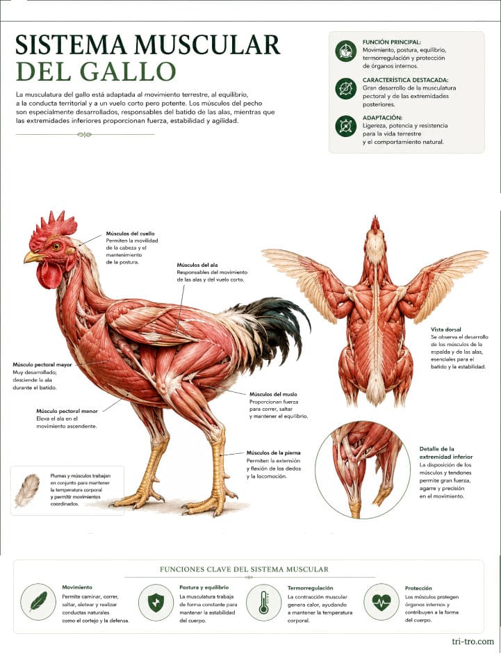
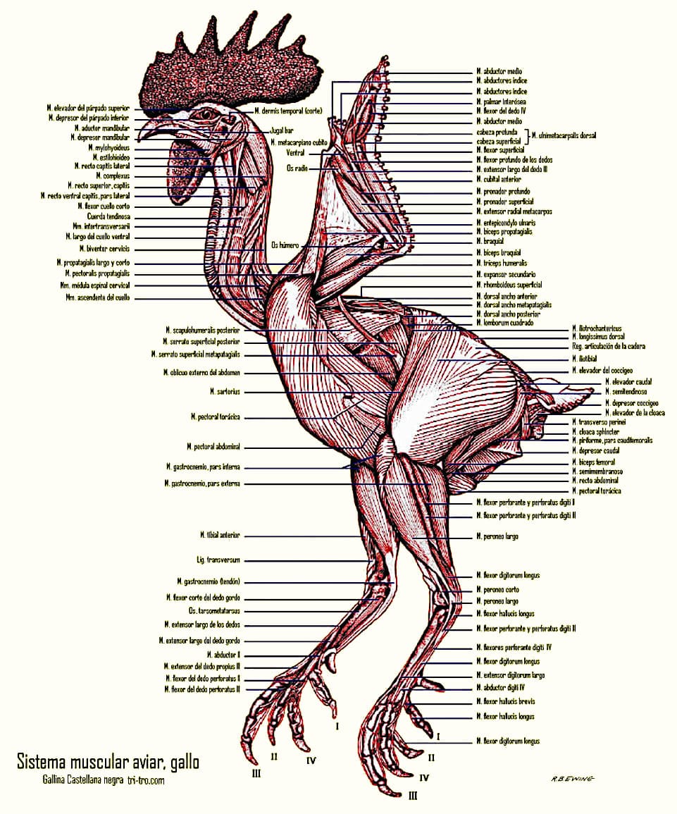
The avian muscular system, in the rooster

Muscle contraction

Muscle contraction occurs in response to a stimulus; it generally originates in the nervous system, either voluntarily or involuntarily. 

When a muscle contracts, the myofibrillar segments shorten as a result of the layers of actin filaments (globular proteins that form microfilaments) and the sliding of myosin filaments (fibrous proteins) along them. The bands running through the myofibril create the striped appearance of skeletal muscle. 

The muscular system provides energy for the bird, and it is here that much of the energy contained in the diet is utilized during involuntary, normal, and voluntary activity.

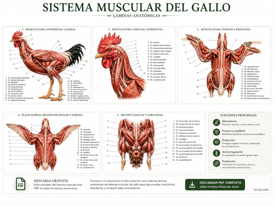
Here is the link to download in PDF the picture of the muscular system bird, rooster.

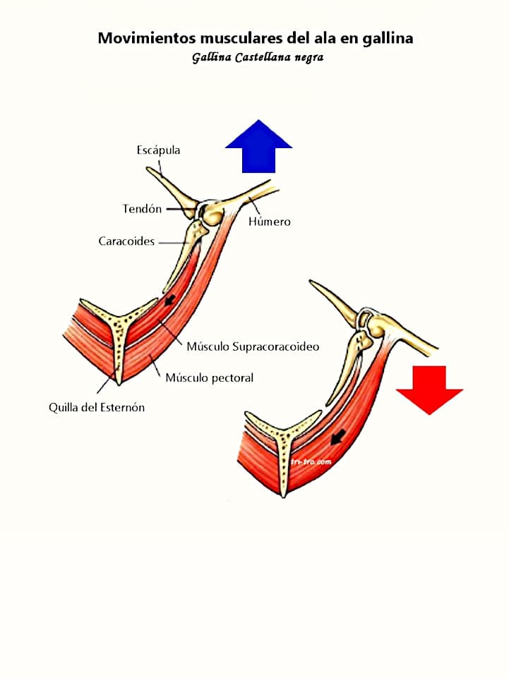
Muscle movements of the wing in hen

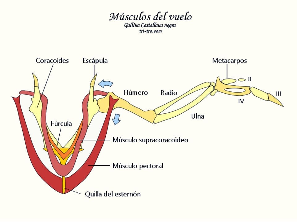
The muscles that move the wings

In the birds that flyare attached to the keel of the sternum, which also holds the vital organs of the abdominal cavity.

Muscle development has increased through genetic selection, as is evident in modern breeds of chickens, broilers, and turkeys raised for meat, although these domestic birds can only fly short distances.

In flying birds, the pectoral muscles are very red, indicating a large number of muscle fibers rich in myoglobin (aerobic oxidative metabolism). Birds with white and red muscles produce white and dark meat, respectively. Red meat contains higher amounts of fat and myoglobin; the latter is a compound that transports oxygen and iron, similar to hemoglobin.

While in the birds that have lost the ability to fly, the musculature is pale, due to the prevalence of muscle fibers white glycolytic (anaerobic metabolism).

It is worth noting the muscles involved in flight; these are the pectoral muscles, which make up the cut of meat commonly known as the “breast.” Generally, these are described as the superficial pectoral muscle and the deep pectoral muscle, also known as the pectoralis major and pectoralis minor, respectively.

The pectoralis major originates from most of the surface of the sternal body, the clavicle, and the sternoclavicular membrane, and inserts into the proximal end of the humerus (pectoral crest). It acts as a wing-lowering muscle during flight.

The pectoral muscle deep originates in the part of the keel sternal, covered by the previous one; it develops a tendon that passes through the channel trióseo to be inserted into the dorsal surface of the proximal end of the humerus. Acts as an elevator of the wing during the flight.

This tendon can break due to trauma (shock or strike it against a window, for example) and then it is characteristic to note that the bird cannot pick up the wing.

In general, the color of a muscle is determined by its activity; red-fiber muscles are the most heavily used; they receive more blood and contain more fat.

White-fiber muscles are rich in glycogen (a white, amorphous substance found in abundance in muscles that can be converted into glucose when the body needs it), which is easily broken down under anaerobic conditions (physical exercise involving short bursts of strength-based activity).

In poultry, the muscles in a chicken's legs are darker than those in the breast, due to the constant effort these muscles exert to keep the body upright when the bird is standing.

In contrast, in non-domesticated flying birds, the breast muscle is darker and serves as support during flight. Broiler chickens have muscle fibers that are larger in diameter and lighter in color than those of our laying hens.

The pectoralis muscles should always be explored, since it indicates the nutritional status of the bird, and can be used for the application of intramuscular injections. It is recommended to introduce the needle into the caudal part of the pectoral muscle surface, by the fact that the head presents a greater blood supply and there is a risk of introducing substances into the bloodstream.

Among the muscles of the forearm, the radial wrist extensor deserves special mention. Originating at the medial epicondyle of the humerus, its short tendon extends over the dorsal surface of the carpal joint and inserts into the extensor process of the metacarpal bone. This tendon can be severed on one wing to prevent flight, as this limits wing extension.

In the hen’s forelimb, in addition to the muscles, it is worth noting the presence of the propatagium or patagium. This is a triangular fold of skin on the cranial side of the wing, extending between the shoulder and carpal joints. Its cranial edge is traversed by the elastic propatagial ligament. To prevent flight, a triangular section of this fold is sometimes removed. It is also used to attach the staples that secure leg bands on waterfowl.

The ulnar cutaneous vein may be visible on the ventral surface; it is suitable for venipuncture (injections and punctures) as it runs over the elbow joint.


Flight muscles of birds

Muscles of the pelvic limb in the chicken

To indicate that your function the main is to keep the body upright and in balance, in addition to being of service to locomotion, so that all the muscles are well-developed.

Can be used to perform intramuscular injections, while always considering that the substances administered in transit through the kidney before they enter the systemic circulation, to exist in birds, a venous system porta-renal.

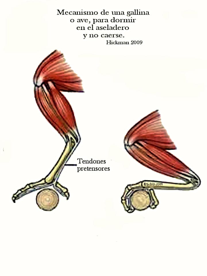
It should also be noted that many perching and prey birds possess what is known as the “reciprocal apparatus.” This system causes the interphalangeal joints of the toes to flex whenever the tarsal joint is also flexed.

This is because the tendons of the digital flexor muscles tighten as they pass over the ventral surface of the tarsus when the bird crouches down (as in the case of a hen sleeping on a perch), causing the toe joints to flex in turn.

This mechanism should be taken into account for desasir the bird, as she jumps of the hanger to let go of the aseladero.

A mechanism that locks the bird's toes to prevent it from slipping off its perch while it sleeps.

On the medial side of the joint the tarsus can recognize the vein of tibial flow. Your journey subcutaneous is ideal for venipuntura (injection and puncture) in birds of a certain size.

Also for diagnostic purposes, as for example in the Marek's disease, it is interesting to know to locate the sciatic nerve in the thigh, caudal to the femur and covered by the muscles medial to the member.

Finally, it should be noted that mineralization of the flexor tendons of the toes (gastrocnemius muscle, superficial and deep digital flexors) and the toes is common; radiographically, this should be recognized as a normal finding.

The muscles of the trunk have a minor importance and the neck are very developed due to the mobility of this region of the rachis. The abdominal muscles and intercostal are reduced to thin films.

In addition, because nearly all of the thoracic and lumbar vertebrae have fused together, the back muscles are less necessary, causing them to shrink in size.

Stiffness cadaveric: When a chicken or other animal dies, the characteristic energy changes are irreversible, and the muscles contract and do not relax for several hours. 

The phenomenon of muscle contraction intensifying is called rigor mortis. 

This condition progresses more rapidly in warmer temperatures or if the animal has suffocated. Muscle relaxation generally occurs 10–12 hours after the onset of rigor mortis due to subsequent changes in the body.

Gallus domesticus Muscle Atlas.

Anatomical Atlas of Avian Muscles

Below is a collection of highly educational anatomical muscle diagrams used to study the musculature of domestic chickens and roosters.

  • Dorsal musculature of the trunk of Gallus gallus domesticus.
  • Superficial muscles of the trunk, viewed from the ventral side, and part of the limbs of Gallus gallus domesticus.
  • Superficial musculature of the left forelimb, lateral view, Gallus gallus domesticus.
  • Deep muscles of the forelimb, lateral view, Gallus gallus domesticus.
  • Superficial muscles of the trunk, lateral view. Gallus gallus domesticus.
  • Superficial cervical muscles of Gallus gallus domesticus.
  • Deep thoracic musculature of Gallus gallus domesticus.
  • Muscles: Cross-section of the right leg and metatarsus at the mid-third of Gallus domesticus.
  • Superficial musculature of the right limb, lateral view, Gallus gallus domesticus.
  • Deep musculature of the right hind limb of Gallus gallus domesticus.
  • Muscles of the cloacal and subcaudal regions of Gallus gallus domesticus.
  • Superficial and intermediate muscles of the thoracic region, dorsal view of Gallus gallus domesticus.
  • Anterior-lateral view of the musculature of the cervical region of Gallus gallus domesticus.
  • Deep cervical muscles of Gallus gallus domesticus.
  • Muscles of the superficial section of the right forearm in its middle third, distal and proximal surfaces, Gallus gallus domesticus.
  • Muscles: Cross-section of the right thigh through its middle third, proximal surface, Gallus gallus domesticus.

These sixteen plates, which will surely be very useful to you. The work was written by Rafael Martín Roldán and published in 1980 by Laboratorios Reunidos S.A.

He holds a degree in Veterinary Medicine from the University of Córdoba and has been a tenured professor of Anatomy at the Faculty of Veterinary Medicine in Madrid since July 17, 1962, having earned the position through competitive examination. He is a full member of the European Association of Veterinary Anatomists, the World Association of Veterinary Anatomists, and the Spanish Anatomical Society, among others. His work includes 25 publications on anatomical and embryological topics related to mammals and poultry. 

Here is the link to download in PDF for all the beautiful illustrations.

Historical anatomical material digitally restored and adapted for TRI-TRO.

Bibliography:

Dyce, K. M., Sack, W. O., and Wensing, C. J. G., 1991. Veterinary Anatomy. Ed. Pan-American.

Ede, D. A., 1965. Anatomy of birds. Ed. Acribia.

Grasse, P. P., 1980. Zoology. Vol. 4: Vertebrates. Reproduction, biology, evolution, and systematics. Ed. Toray-Masson.

Grifols, J. and Molina, R. 1994. Manual of clinical exotic birds. Ed. Grass-Iatros.

Natural History, 1984. Vol. 1. Ed. Ocean-Institute Gallach.

King, G. M. and D. Custance. Colour atlas of vertebrate anatomy: The Pigeon. Pp.: 5.1— 5.17. Ed. Blackwell Scientific Publications.

Krahmer, R., and Schröeder, L. 1979. Anatomy of the domestic animals. Ed. Acribia.

McClelland, J., 1992. Atlas in color of the anatomy of birds. Ed. inter-American.

Laboratories Meeting SA Anatomy of the hen D. Rafael Martin Roldan.

Nickel, R., 1977: The Anatomy of the Domestic Birds. Ed. Paul Parey.

Sandoval, J., 1976. Veterinary Anatomy. Ed. Modern Printing Press (Córdoba).

Schwarze E. 1980. Compendium of Veterinary Anatomy. Volume V: Anatomy of birds. Ed. Acribia.

Shively, M. J., 1993. Veterinary anatomy, basic, comparative, and clinical. Ed. Manual Moderno.

Sturkie PD, 1967. Physiology of Avian. Ed. Acribia.

Dr. Francisco Gil Cano.

Professor H. Aguinaga.


You may be interested in these other sections


The Rooster's Reproductive System: Parts and Function

Gallus lafayettii: Origin and Characteristics of the Ceylon Fowl

Exploring the Anatomy of the Domestic Rooster


Do you want to share or comment on your social networks

Scan to visit TRI-TRO
Escanea para visitar. TRI-TRO