Parasitic diseases:
Syngamus trachea, conocido también como The tracheal worm is a roundworm that, as its name suggests, is found in the trachea of various types of poultry.
Birds (whether adults or young) that show no other signs of illness yawn constantly; in reality, however, they are trying to get more air because they feel suffocated or short of breath when their airways are constricted by worms, and they may even make a motion as if to yawn, as if trying to expel something from their throats.
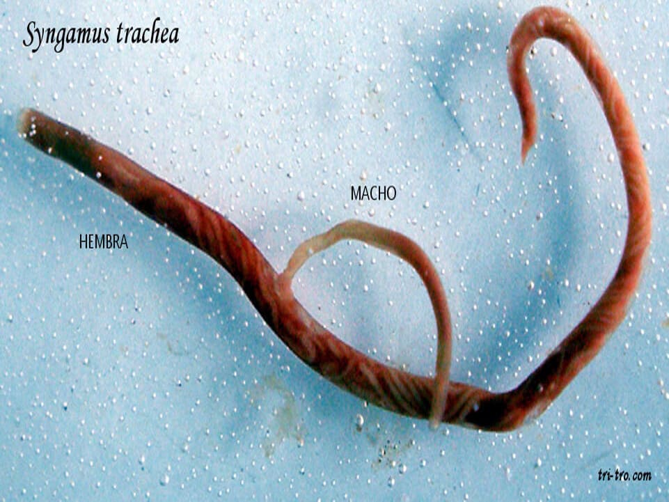
This parasite can cause problems, ranging from severe shortness of breath until the possible death of the animal. Both parasites, male and female, they remain united in intercourse life.
The singamosis Yawning disease is a parasitic disease characterized by the presence of nematodes in the upper respiratory tract, trachea, and, rarely, the bronchi, caused by the Syngamus trachea.
Birds (whether adults or young), showing no other signs of illness, yawn constantly—though in reality they are simply trying to get more air because they feel suffocated—or even make a motion as if to yawn, as if trying to expel something from their throats.
Also related to the tracheal worm, a roundworm that, as its name suggests, is found in the upper respiratory tract—specifically, the trachea—of various types of birds. This parasitic disease typically occurs at the start of summer and can affect all domestic birds, but Syngamus trachealis shows a preference for pheasants, which include chickens, pigeons, etc.
It has been observed that this causes problems, ranging from severe breathing difficulties to, in some cases, the death of the bird.
I'm sure we've all come across a hen or rooster on more than one occasion that snores slightly when breathing, shakes its head for no apparent reason, or coughs.
The presence of worms in the respiratory system causes more severe symptoms in young birds, characterized by difficulty breathing, loss of appetite, general lethargy, and weight loss.
These small nematodes (roundworms) are hematophagous (meaning they feed on blood). Once they have entered the body of a bird, such as a chicken (the final host), the parasite’s preferred sites are usually the trachea and bronchi; they are rarely found in the lungs or air sacs. There, they attach themselves firmly to the epithelium and pierce it, feeding on the blood supplying the area.
In life of the parasite, its coloration shows a tone is bright red, due to the abundance of blood in your digestive system.
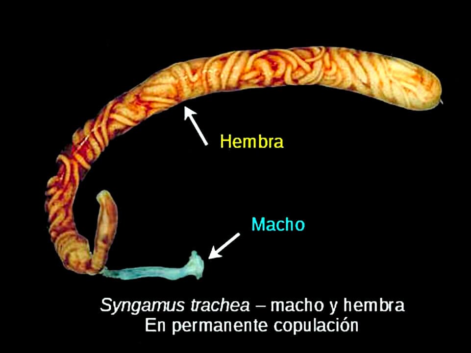
The female, of two inches in length, with the vulva located very close to the anterior end, where the male is attached by the structures found on his posterior end, known as spicules; these are accessory genital organs used for penetration, located at the tip of the male nematode’s tail, near the copulatory sac, known as the copulatory pouch.
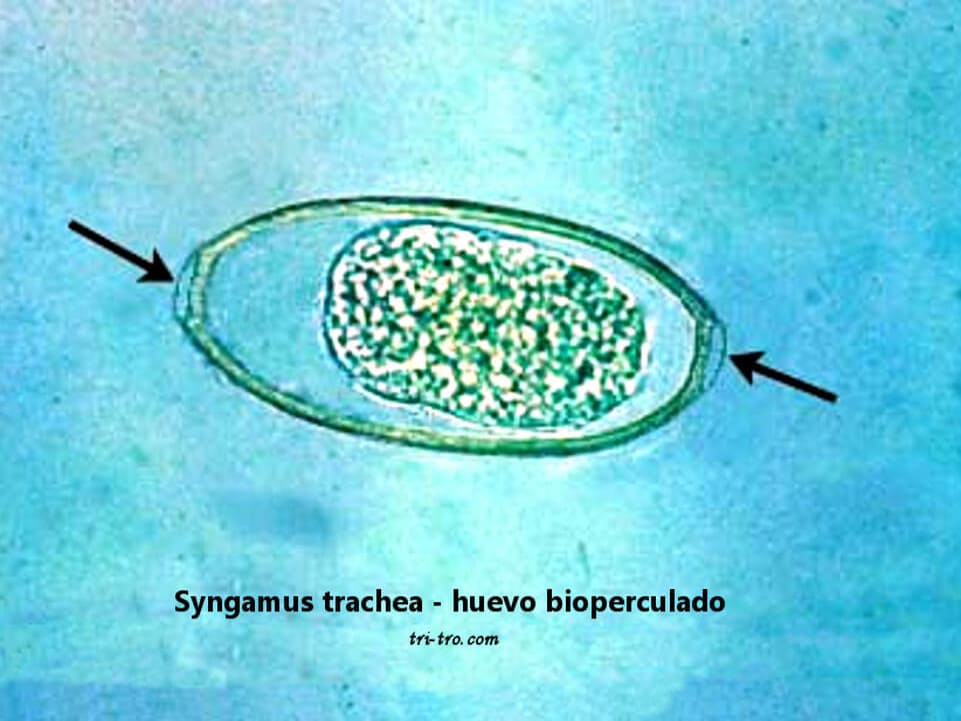
The male, measuring 0.6 centimeters, is considerably smaller than the female. The parasite's eggs are shaped like lemons, with two opercula at each end. Both el macho como la hembra estarán unidos en cópula de por vida, tomando la forma de un tenedor, color rojo por la abundante sangre en su interior.
Constant copulation is one of the most striking characteristics of this species, giving them a fork-like appearance—hence one of their common names in Spanish, “gusano horquilla” (fork worm).
The mouth with which the parasite attaches itself to the epithelial wall to draw out and suck blood is cup-shaped; this distinctive feature is the source of one of its English names, the gapeworm.
The female lays eggs that will be expelled by the bird, along with tracheal mucus secretions, through coughing and sneezing, or in feces.
Thanks to this type of reproduction (continuous mating), egg production is constant. The eggs are elliptical in shape and measure approximately 45×80 µm (microns); they are covered by a thick cuticle, enabling them to withstand various environmental conditions, and have an operculum at each end.
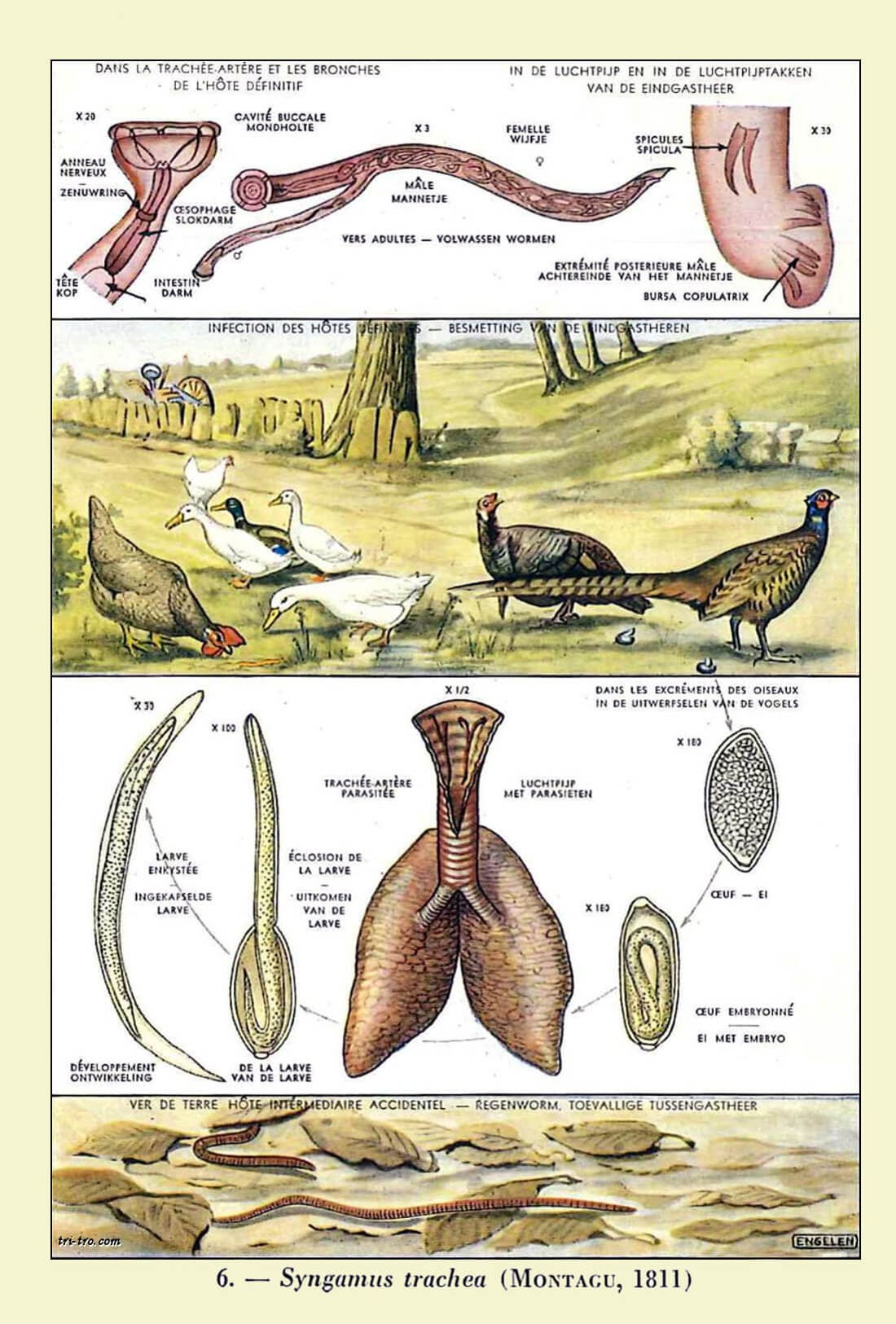
Once outside the host, where it finds optimal temperature and humidity conditions, the egg—still in its embryonic stage—will go through three stages of development, reaching the final stage between 8 and 16 days after the female lays it. This is how they infest birds, by entering their digestive system with food. If the egg is not ingested by the bird, the larva will hatch and will be ready to infest the bird in the same way.
Earthworms, snails, slugs, and small beetles, etc., are the ones that eat the eggs and larvae of the Syngamus trachea, and then disseminate them in faraway places, thus increasing the radius of action for the contamination of the birds.
Once the bird has ingested both the eggs and the larvae, they travel through the digestive tract; in the upper sections of the intestine, the larvae penetrate the intestinal lining to reach the fine capillaries, through which they travel to the liver, heart, and finally the lungs.
A massive infestation of larvae in the lungs can lead to severe pneumonia, especially in young animals; the parasites reach the bronchi.
There, they will molt twice, reaching adulthood in six days after being ingested by the bird; males and females will pair for life, then travel to the trachea, where they will settle in their final location, and within two weeks, the female will begin laying eggs, thus initiating the parasite’s life cycle.

In more advanced cases, respiratory symptoms appear, including coughing fits (a reflex that triggers coughing when the efferent nerves of the trachea are stimulated) and the expulsion of mucus through the nostrils and the beak.
To confirm its presence and directly observe the adult parasite in the trachea, one can use a strong backlight, remove the neck feathers, and stretch the exposed skin. Endoscopy can also be used. However, the easiest way is to detect the presence of eggs using a microscope:
The Syngamus trachea can be transmitted to our chickens direct as well as host.
Direct:
By the ingestion of the hen, with the food in the feeders, water, stained or with traces of feces of infected birds, the remains of mucus tracheal infected, between ave and ave, reached the mouth when coughing, or by pecking substrate contaminated with eggs or larvae, in the courtyard of the chicken coop or surroundings, garden.
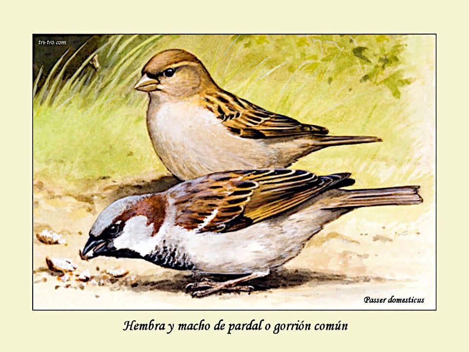
A female and a male house sparrow. Passer domesticus
Hosts:
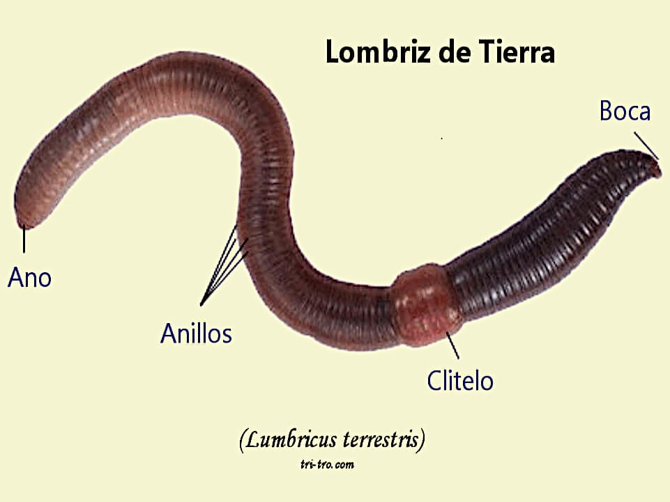
The eggs of the parasite Syngamus trachea, as well as their larvae, are found outdoors and can be ingested by a wide variety of animals, such as house sparrows, doves, and thrushes, which then become carriers or vectors of the infection. In the soil, earthworms, snails, slugs, and even insects, such as in the case of the flies.
Some studies suggest that the infectivity of the larvae of Syngamus trachea It tends to increase when its life cycle involves a host, especially in the case of earthworms.
Earthworm, lumbricus terrestris
The larvae, when you have an intermediate host, interrupts their growth and is enquistan within the tissues of the animal. Many times in areas of easy access for predators.
These cysts can remain viable, and, therefore, ineffective, and for extended periods of time.
The duration of the parasitic infection phase ranges from 12 to 14 days. Certain larvae, migrating directly through the intestine, are able to reach the lungs by traveling through the peritoneal cavity (a very thin space located between the two layers of the peritoneum: the parietal layer and the visceral layer), which prolongs the incubation period and delays the onset of symptoms in the hen.
This parasite is transmitted through oral ingestion, so the primary measures will focus on controlling food, grit, and water.
Its control is fairly simple, and still have not been documented serious problems of resistance to anthelmintics normal part of the Syngamus trachea.
Detected the problem, your veterinarian will be able to advise you about the type of drug used, its dose and time of administration.
The most commonly used drugs—levamisole (Ergamisol), fenbendazole (Panacur), and ivermectin—can be effective in treating resistant strains.
In addition to treatment, it is important to implement a plan to reduce the risk of infection by limiting the hens’ exposure to sources of infection, preventing wild birds from entering chicken coops or yards by installing mesh screens they cannot pass through, and maintaining strict hygiene standards for feeders and waterers.
Literature review:
MERCK & CO. (1995). Manual Merck de Veterinaria. Rahway, N. J., EE. UU.
BUXADÉ, P. (1987). The laying hen. Ed. Mundiprensa. Madrid.
DORN, P. (1987). Manual of avian pathology. Ed. Acribia. Zaragoza.
HOFSTAD, M. S. (1984). Diseases of Poultry. Iowa State University Press, Ames, Iowa.
ZARZUELO, E. (1982). Vade mecum of the pathology, infectious poultry. Ed. Aedos, Barcelona.
CASTELLÓ, F. and CASTELLÓ, J. A. (1960). The New Art of Raising Chickens. Aedos, Barcelona.
OROZCO, F. (1989). Breeds of chickens Spanish. Ed. Mundiprensa. Madrid.
LACADENA, J. R. (1998). Genetics. Ed. AGESA
PUERTAS, M.J. (1992). Genetics: Fundamentals and Perspectives. McGraw-Hill Interamericana.
SANCHEZ-MONGE, E. (1969), Genetics. Espasa-Calpe S.A.
OROZCO, F. and ROBLA, F. (1986). Genetic aspects of the León rooster. 24th Symposium of the WPSA (Spanish Section): 199–212.
HILL, J. L. (1973). Genetics, general and applied. Ed. UTEHA.
CASTELLÓ, J. A., LLEONART, R., FIELD, J. L., OROZCO, F. (1989). Biology of the chicken. Real Escuela de Avicultura.
LLEONART, F., ROCA, E., CALLÍS, M., GURRI, A., PONTES, M. (1991). Poultry Hygiene and Pathology. Royal School of Poultry Science.
STURKIE, P.D. (1968). Fisiología aviar, ed. Acribia. Zaragoza.
LOHMANN ANIMAL HEALTH (2012)
Paul Junquera, 2013, Parasitipedia.net.
Robert B. Altman, Susan L. Clubb, Gerry M. Dorrestein and Katherine Quesenberry, 1997, Avian medicine and surgery.
Lapage G, 1968, Veterinary Parasitology.
Kaufmann, J., 1996, Parasitic Infections of Domestic Animals.
Hieppe, T., 1985, Lehrbuch der Parasitologie. Band 3. Veterinärmedizinische Helminthologie.
You may be interested in these other sections