Parasitic diseases:
Trichomoniasis is a disease a parasitic disease that affects various birds and chickens in the upper part of their digestive tract and is caused by a flagellated protozoan known as Trichomona gallinae.
The disease has a chronic course, with Trichomonas spreading to the oral cavity, sinuses, pharynx, crop, and sometimes even the proventriculus.
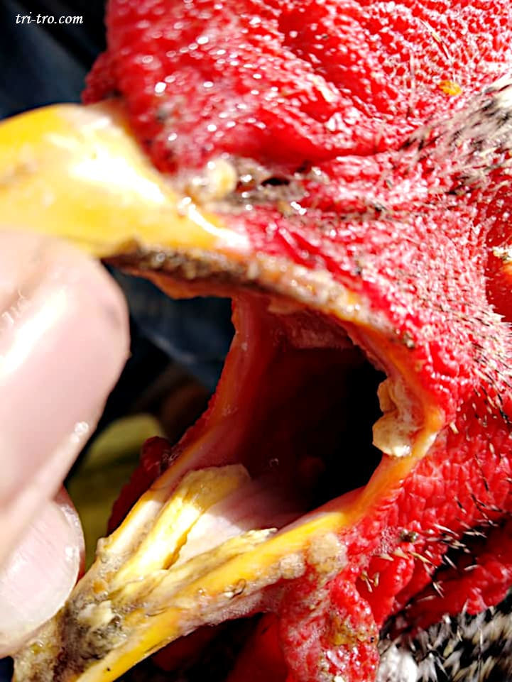
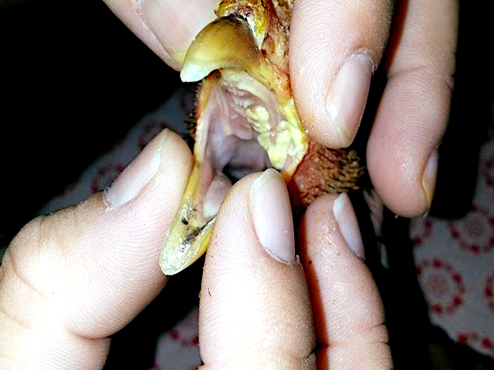
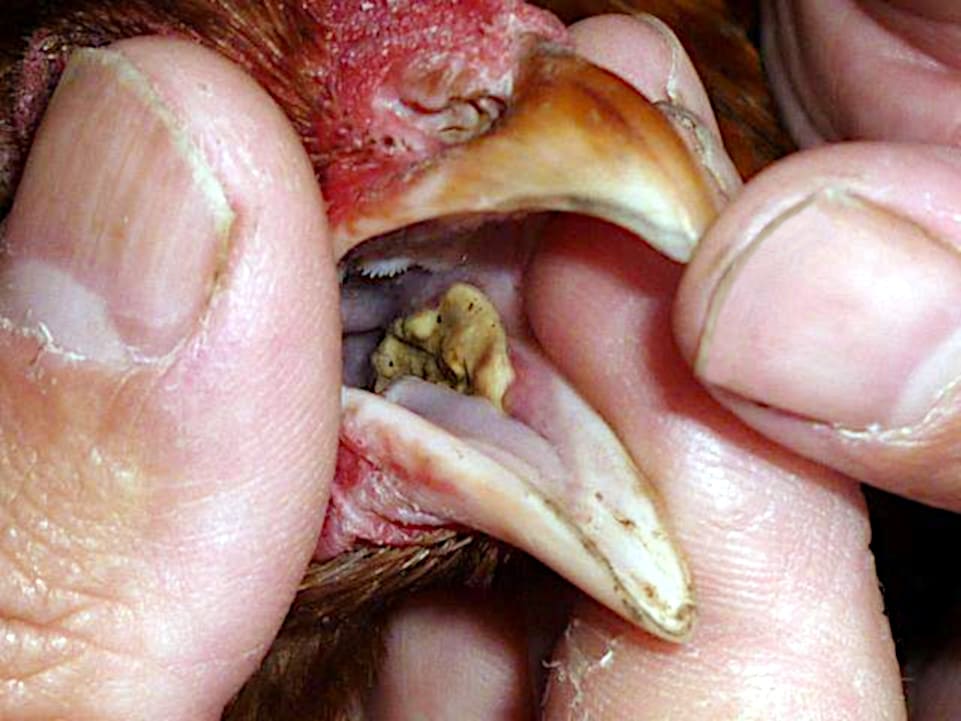
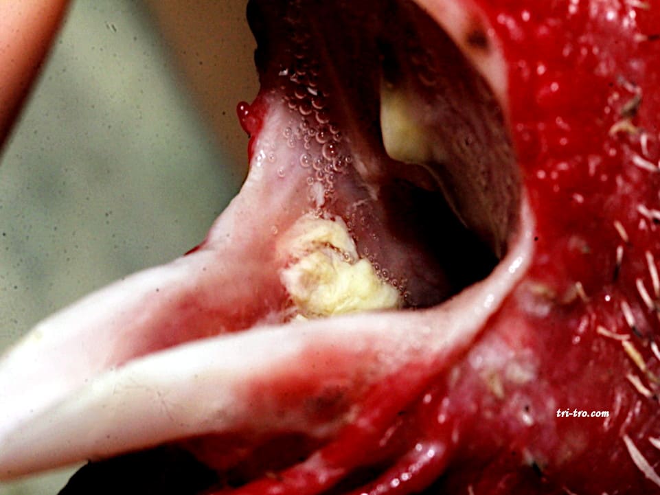
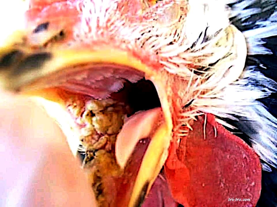
The lesions consist of a mass casposa surface yellowish, forming ulcers.
Birds affected present with cyanosis, hypertrophy, sinus, and arching of the neck.
It is mandatory to cull the most severely affected animals, as they are permanent carriers; in addition, strict hygiene measures must be taken regarding drinking water.
Gastrointestinal parasites can cause many problems in a bird’s stomach and intestines, but they also affect the normal functions of other organs. One such parasitic infection is trichomoniasis.
Trichomoniasis, also known as canker or sores, is an infection caused by Trichomonas gallinae, a protozoan (or single-celled microbe). It generally affects wild birds and is occasionally seen in domestic birds.
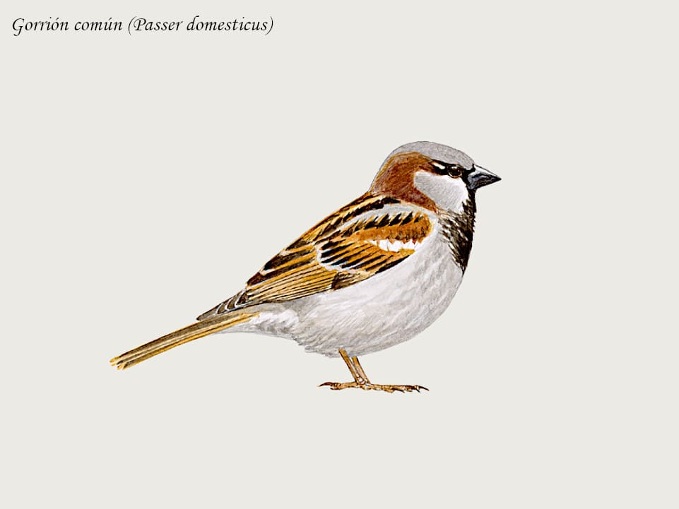
Trichomoniasis is a parasitic disease caused by the protozoan flogging Trichomonas gallinae it primarily affects birds Columbiformes and birds of prey, being occasional, galliformes, passerines and psitaciformes (Gross, 1996).
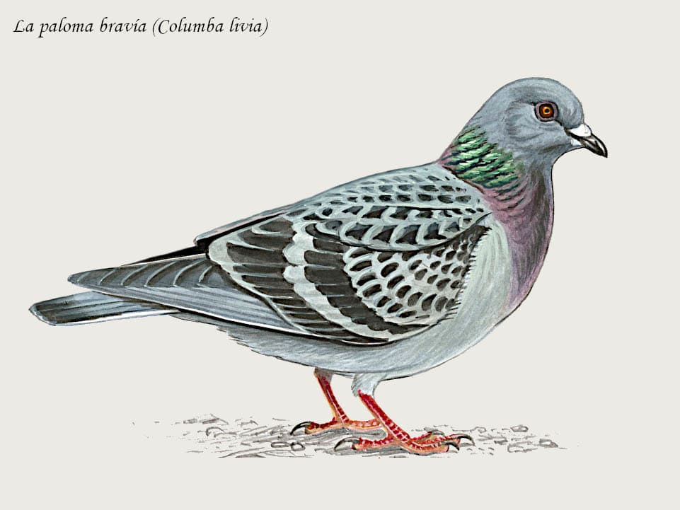
Are the main guests of Trichomonas gallinae; almost all pigeons are carriers, although small outbreaks of the disease sometimes occur. The disease is more severe in young birds (affecting them more easily and aggressively), and adult birds may be carriers of the disease, whether symptomatic or not (asymptomatic carriers, meaning they have the protozoa but show no symptoms of the disease).
Trichomoniasis in pigeons is transmitted from adult birds to their chicks during feeding (regurgitation), infecting the chicks within minutes of hatching. Infection with T. gallinae may be asymptomatic or symptomatic, and can progress to fatal cases within 4 to 18 hours of infection.
Trichomoniasis is considered the main cause of death among the pigeons newborn. The hawks and other wild birds can become infected after feeding on an infected bird.
In our poultry houses, hens, chickens, and turkeys become infected with the protozoan parasite through water contaminated by wild birds and through feed or grain, following contact with waterers and feeders.
The trophozoites of Trichomonas gallinae can stay alive for several hours in the water and during 5 days in the grains, and are capable of infecting other birds that feed in these troughs (Borges, 2006).

The course of the disease is rapid. Early lesions appear as small areas of yellow in the oral mucosa. Increase in size rapidly and fuse to form masses that end up blocking the esophagus, and can prevent the bird to close its beak. Fluid may build up in the peak.
It produces a watery discharge from the eye and, in the latter stages, an exudate around the eyes can cause blindness. The birds lose weight quickly and become weak and apathetic. Death occurs within 8 to 10 days. In chronic infections, the birds seem to be healthy, although parasites can usually be demonstrated in those that scrape the mucous membranes of the throat (Fraser et al., 1996).
In addition, the bird can be full of foci of necrotic home. The peak and the esophagus contain material necrotic, which may extend to the inside of the skull and sometimes through the surrounding tissues of the neck to wrap around the skin. In the esophagus and into the crop, the injuries can be yellow, rounded, and high, with a spur chestnut known as “yellow buttons”.
The crop may be covered by a yellowish diphtheritic membrane that can extend into the glandular stomach. However, the gizzard and intestine are not affected. Damage to internal organs is most commonly seen in the liver. Adhesions and involvement of other internal organs appear to be contact extensions of the liver lesions, according to Fraser et al., 1996.
In the throat, we must distinguish trichomoniasis from other diseases that cause similar lesions, including
Viruela: The plates are not located at the back of the throat, but rather on the inner sides of the beak; they are very difficult to remove.
The trick to telling them apart is to use a cotton swab—always while wearing gloves—and gently remove a small amount of the yellowish plaque; if it bleeds heavily, it’s diphtheria-smallpox; if it bleeds only a little, it’s trichomoniasis.
Mycosis – Fungal Infections – Candidiasis: These appear as yellow spots on the sides of the palatal fossa or opening above the palate; they are not caseous.
The most important thing to do in our chicken coops if we notice that our birds are infected with trichomoniasis is to wash the equipment used for feeding (feeders, feed buckets, egg collection baskets, etc.) and for water (primarily drinkers and water troughs) frequently and thoroughly, thereby preventing the spread of the disease.
Keep the coop clean at all times and clean perches, plates, walls, doors, windows, etc., with frequency.
Prevent wild birds from entering the chicken coops; this is very important.
Seeing that they are sick of our birds, the best procedure is to seek the help of a veterinarian for a proper diagnosis and treatment.
Medications for trichomoniasis are drugs derived from nitroimidazoles. They are effective against trichomonads, amoebae, Giardia, histomonads, and bacteria.
Metronidazole: It is a heterocyclic compound with a chemical structure similar to that of nitrofurans. It is commonly used under the brand name Flagyl. Among the most recent trichomonicides to enter the market are carnidazole, sold under the brand name Spartrix, and ronidazole. Ideally, different trichomonicides should be rotated.
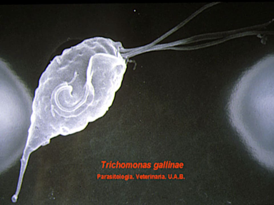
trichomonas gallinae
Trichomonas gallinae: The The cell has a pear-shaped body and consists of cytoplasm and a nucleus, which is located at the front of the cell.
The basal bodies are located on the front edge of the body, from which 3–4 flagella extend forward and one extends backward along the membrane.
With the help of flagella and membrane, trichomonas move in rotation about the longitudinal axis of the body forward. This explains the movement of the membrane-left and right.
Trichomoniasis avian
Literature review:
MERCK & CO. (1995). Manual Merck de Veterinaria. Rahway, N. J., EE. UU.
BUXADÉ, P. (1987). The laying hen. Ed. Mundiprensa. Madrid.
DORN, P. (1987). Manual of avian pathology. Ed. Acribia. Zaragoza.
HOFSTAD, M. S. (1984). Diseases of Poultry. Iowa State University Press, Ames, Iowa.
ZARZUELO, E. (1982). Vade mecum of the pathology, infectious poultry. Ed. Aedos, Barcelona.
CASTELLÓ, F. and CASTELLÓ, J. A. (1960). The New Art of Raising Chickens. Aedos, Barcelona.
OROZCO, F. (1989). Breeds of chickens Spanish. Ed. Mundiprensa. Madrid.
LACADENA, J. R. (1998). Genetics. Ed. AGESA
PUERTAS, M.J. (1992). Genetics: Fundamentals and Perspectives. McGraw-Hill Interamericana.
SANCHEZ-MONGE, E. (1969), Genetics. Espasa-Calpe S.A.
OROZCO, F. and ROBLA, F. (1986). Genetic aspects of the León rooster. 24th Symposium of the WPSA (Spanish Section): 199–212.
HILL, J. L. (1973). Genetics, general and applied. Ed. UTEHA.
CASTELLÓ, J. A., LLEONART, R., FIELD, J. L., OROZCO, F. (1989). Biology of the chicken. Real Escuela de Avicultura.
LLEONART, F., ROCA, E., CALLÍS, M., GURRI, A., PONTES, M. (1991). Poultry Hygiene and Pathology. Royal School of Poultry Science.
STURKIE, P.D. (1968). Fisiología aviar, ed. Acribia. Zaragoza.
LOHMANN ANIMAL HEALTH (2012)
You may be interested in these other sections