Poop apparently normal of chicken, which are those little white dots
Here increased, being perfectly Tapeworm larvae
This topic can be a little unpleasant For some of you: if you have a bit of a weak stomach or find the analysis of your birds’ foul-smelling droppings tedious and disgusting, I suggest you move on to other topics.
As for the rest, let’s take a look at the intestines—what parts they’re made of, defecation, feces, or droppings, their composition, color, and whether they’re hard or soft—of our beloved chickens.
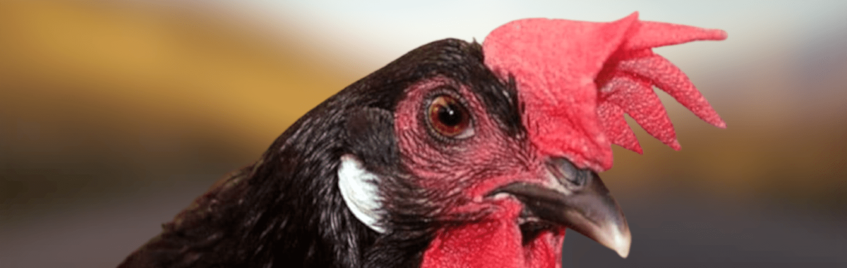
We all know a bird is sick when it’s withdrawn and keeps to itself, seems depressed, has ruffled feathers, isn’t eating, etc.
Breeders' top priority is always the health of their chickens or other birds. One of the most important ways to monitor their health is to observe and examine their droppings, which can indicate whether they are normal or suggest the presence of any diseases, whether gastrointestinal or parasitic.
The problem is that this description of their droppings alone is just a small piece of the puzzle when it comes to diagnosing what’s wrong with our chickens.
As you already know, the treatment of the symptoms never works; that is, you must find mainly the cause and then treat.
Often, finding the cause can be as simple as taking a closer look at the fresh droppings from our chickens or other birds. Color se ve influenciado por the diet ave.
What is all this from the droppings?
Bird droppings are quite distinctive because all of the waste is expelled at once, in a small, neat, bubble-like or tube-shaped form.
Normal stool is cylindrical in shape, reflecting the size and diameter of the intestine.
Together with the stool, it is a variable amount of uric acid or urate (cal) and urine (water). The urates are usually in the form a bubble or mixed in with the stool and should be white or beige.
Chicken droppings consist of three parts:
They are a byproduct of the kidneys and are usually snow-white when dry. They consist of the crystalline portion of urine (uric acid) and have a consistency that is neither very liquid nor very solid. Urates with yellow or green discoloration are a sign of trouble.
It's the clear part, and it's like water; in fact, it's not much different from any of the others animals. Sometimes, urine and urate crystals mix together to form a cloudy liquid; there is no need to be alarmed if you do not see the two distinct areas.
This is the third part, and it’s the only one that’s actually solid. It’s that section in the middle that looks like a tube. It can be straight, spiral-shaped, or even broken into smaller pieces, but it still retains its tubular shape.
Stools can be normal or abnormal:
It’s important to keep in mind that it’s normal to see droppings when our birds are healthy. This is because healthy birds eat a rich and varied diet, and their coops, feeders, waterers, and, of course, their yards are kept very clean.
When they get sick, the droppings will always be abnormal, either by the color and consistency, odor, etc. These observations of the stool are what can indicate health problems.
Always remember that the diet, stress, and the environment will have a natural effect about the droppings of the birds.
During times of stress, droppings will naturally be more watery; this is mainly because, when a “fight or flight” response occurs, this bodily process in birds is triggered by an instinctive reaction.
When the birds are scared, they expel the waste out of your system before taking flight, or flee. These cacas will have more amount of urine, and urates in the feces each day.
If the stress is ongoing, most droppings will contain very little feces, or none at all.
Also, you will notice plenty more piss in the feces if a bird drink in excess or eats food with high content of water. Lettuce and fruit have a very high water content and are the most common cause of watery stools.
The color of the stool may change from time to time, depending on what the chicken has eaten recently.
Seeds Green vegetables naturally produce green droppings, while blueberries and blackberries produce black droppings; similarly, if they have eaten ash or charcoal mixed with sand to deworm themselves, their droppings will also be black.
If we feed our hens with pellets in the diet, the stools tend to be the same color as the pellet, if these are colored or rusty color or if they are colored.
If we see that the stool of our birds are bad, we will keep the diet simple, that is to say, re-seeds, for a few days and come back to check.
Fortunately, our chickens' digestive system works very quickly, and they won't have any foreign matter in their system, since they expel it from their bodies relatively quickly.
We must always keep this in mind and pay close attention to our birds’ droppings in order to identify segments of worms, the eggs and larvae of parasites, from the respiratory system, digestive tract, digestive system and excretory.
You are interested in this section, hierarchy in the chicken coop, since they are gregarious and prone to stress; they need to feel at ease within their group.
In the step of the food there is the presence of particles does not digested food by the feces of poultry and causes a decrease in the digestive efficiency with economic consequences (feed conversion, growth, productivity of the channel, the costs of production, etc.).
Diarrhea:
When a bird has diarrhea, its droppings will be soft and won't have the typical cylindrical shape.
A chicken or bird has diarrhea when its feces are loose and formless. Diarrhea is not very common in birds. A drooling condition with normal feces but a large amount of urine around them is known as watery drooling (polyuria); this should not be confused with diarrhea.
Diarrheal stools appear loose, but not all loose or watery stools constitute diarrhea. It’s important to keep this in mind; now we’ll look at other forms of diarrhea.
Secretory diarrhea:
This involves excessive secretion of fluid from the intestinal mucosa, which affects the intestine's ability to absorb fluids.
This is caused by the virus that destroy enterocytes mature on the tips of the villi, leaving enterocytes secretory functional in the crypt and on the side of the villus.
It may be caused by bacterial enterotoxins that affect the mediators of intestinal electrolyte transport, involving epithelial cells of the mucosa, immune and mesenchymal cells, enteric neurons, and cells of the endocrine and nervous systems central.
These mediators act by increasing chloride secretion from the crypts and decreasing NaCl absorption at the tips of the villi; as water follows these electrolytes, the net result is fluid overload in the intestinal lumen.
Osmotic diarrhea:
This involves forces of osmotic excessive exerted by solutes luminal.
The diets of poultry with a lot of salt is one of the causes, such as occurs with diets formulated with certain batches of flour by-product of a bakery.
Osmotic factors may be involved in digestive problems associated with antinutritional factors (non-starch polysaccharides, NSPs) in rye, barley, wheat, and other ingredients, as reviewed by Ijl, 1999.
These complex carbohydrates, typically hexoses and pentoses, are resistant to digestive enzymes, creating a viscous environment within the intestinal lumen, increasing the mass of the luminal digesta, and producing sticky, moist feces.
It’s a good idea to have a basic understanding of the foods we feed our birds and how they benefit them.
Produces bowel movements cacas or bulky stools with increased bulk osmolarity due to unabsorbed nutrients. This occurs because intraluminal digestion is impaired due to ineffective enzymes or a lack thereof.
Malabsorption may occur due to the loss of mature enterocytes, which have been replaced by immature cells that lack full absorptive function. Once again, NSP may be involved in malabsorption because gelatinous luminal blocks prevent enzymes from accessing nutrients that would otherwise be digestible.
This problem is also associated with increased mitotic activity and greater depth of the intestinal mucosal crypts; this suggests increased turnover of enterocytes and a relative increase in secretory enterocytes or a decrease in mature enterocytes. The concentration of bile acids in the digestive contents is also diluted, possibly impairing lipid emulsification.
The concentration of the bile decreased is associated with increases in aerobic bacteria and anaerobic luminal.
Diseases exudate:
In this situation, the intestine undergoes severe damage involving necrosis and the loss of enterocytes, fluids, electrolytes, and plasma from the damaged mucosa, accompanied by a significant inflammatory response.
The host must deal with the escape of pathogens in a primary or secondary in the vascular system and the impact on the liver, and possibly beyond.
Even if the disease itself is not severe, anorexia and the diversion of nutrients toward inflammation and tissue repair will reduce growth and performance, impairing feed conversion and increasing production costs.
Toxic injuries:
When oxidized, rancid fats produce free radicals that can cause sublethal damage to cells throughout the body, including the intestinal epithelium.
The most obvious clinical sign of rancid fats is vitamin E deficiency, which manifests as encephalomalacia.
Biogenic amines, generated by the bacterial decomposition of the fish tampered with and substrates provided, are directed to the tissues gastrointestinal.
Polyuria:
Abnormally large amounts of urine produced by the bird (polyuria) are harder to detect in some birds. This disease is often fatal and has the most severe impact on young chickens.
The few chickens that survive to adulthood develop, often, damage to the plumage and malformations of the beak.
Polyuria can be caused by a viral infection, stress, kidney disease, or poisoning. In our birds, causes include excitement and anxiety.
Urine in the stool may indicate underlying health conditions (such as diabetes or kidney disease), but it is usually caused by increased water intake or large amounts of water-rich fruits and vegetables.
The mucus that is secreted On the epithelial surface, it lubricates the movement of food through the digestive tract. It is secreted by specialized epithelial cells arranged in glands in the mouth and esophagus, and by individual goblet cells in the proventriculus and intestine.
Mucus is not secreted in the crop or gizzard; however, the chyme reaches these organs softened and lubricated by the glands of the mouth and esophagus. Mucus is a viscous substance composed of water and glycoprotein. It protects the cells of the mucosa in the stomach and intestine from self-digestion caused by gastric acid, pepsin, and digestive enzymes.
The protective effect of the mucosa is evidenced by increased secretion on the mucosal surface and hypertrophy of goblet cells in response to harmful stimuli. One of the properties of mucus is that it acts as a barrier against invasion by bacteria and fungi. Virulent strains of Candida albicans, the agent of thrush, they have an enzyme that dissolves (mucinolytic) the barrier of mucin in order to improve the adhesion and penetration of epithelial cells.
Helicobacter pylori, the causative agent of gastric ulcers in humans, exudes urease that breaks down the protective layer of the gastric mucosa.
Poultry feed, soya flour, well heated, may contain urease.
In addition to mucus, the intestine secretes large amounts of water mixed with electrolytes.
It is estimated that, for every gram of food ingested, the intestine secretes approximately 2 grams of water to aid digestion and absorption. Excess water in the lumen is reabsorbed in the lower small intestine, the cecum, and the colon. The fluid in the upper small intestine, however, serves a protective function by keeping bacteria in suspension and flushing them downstream.
Bibliography:
'argenzio, AR. Pathobiology neuroimmune diseases enteric infectious. The mechanisms of the pathogenesis of enteric diseases, ed. Paul, PS, DH Francisco, and GIVES Benfield. Advances in Medicine and Experimental Biology, 1997.
Baba, E, H Wakeshima, K Fukui, et al. The adhesion of bacteria to the mucosal surface cecum of chickens conventional and germ-free, American Journal of Veterinary Research, 53, 1992.
Hill, AR, F, Aumont, and N. Deslauriers, et al. Evidence of degradation of mucin gastrointestinal Candida albicans aspartyl proteinase secretory. Infection and Immunity, 64, 1996.
Copemen, M, J Matuz, AJ, Leonard, et al. The barrier gastroduodenal mucous membrane and its role in the protection against pepsinas luminal: the effect of 16, 16 dimethyl prostaglandin E2, carbopol-polyacrylate, sucralfate, and bismuth subsalicylate. Journal of Gastroenterology and Hepatology, 9 Suppl 1, 1994.
Crawford, JM. The oral cavity and the gastrointestinal tract. In basic Pathology, Sixth Ed. EDS. Kumar, V, RS Cotran, and SL Robbins. WB Saunders Company. 1997.
Elson, CO In defense of mucosal surfaces: The regulation and manipulation of the immune system of the mucosa. The mechanisms of the pathogenesis of enteric diseases, ed. Paul, PS, DH Francisco, and GIVES Benfield. Advances in Medicine and Experimental Biology, 412, 1997.
Hoerr, FJ, WW Carlton, B Yagen. Micotoxicosis caused by a dose singe of toxin T-2 or diacetoxiscirpenol in the diet of broiler chickens. Veterinary Pathology, 18, 1981.
Hofacre, CL, R ROYMAN, B Gautrias, et al. The use of Aviguard and other bioproducts intestinal in experimental Clostridium refractile-enteritis, necrotizing associated in broiler chickens. Avian Diseases 42, 1998.
Iji, PA. The impact of cereal non-starch polysaccharides in the development and intestinal function in broiler chickens. Poultry Science Journal World 55, 1999.
Leeson, S and J. d. Summers. Commercial Poultry Nutrition. 2nd ed. University Books. Guelph, Ontario, Canada. 1997.
Moon HW. Histopathology comparison of infectious intestinal. On the mechanisms of patho-genesis of enteric diseases, ed. Paul, PS, DH Francisco, and GIVES Benfield. Advances in Medicine and Experimental Biology, 1997.
Riddell, C and XM Kong. The influence of diet on necrotic enteritis in broiler chickens. Avian Diseases 36: 499-503, 1992.
Siegel, Pb, As Larsen, CT, Larsen, et al. The resistance of chickens to an outbreak of necrotic enteritis, as influenced by the genotype main chisto-support and fund genome. Poultry Science 72,1993.
Smith, AW, B Chahal, and GL French. The pathogenic human gastric Helicobacter pylori cholerae. Molecular Microbiology, 1994.
Smits, CHM, A Veldman, and HJ Verkkade, et al. The inhibitory effect of carboxymethyl cellulose high viscosity in the absorption of lipids in broiler chickens coincides with a reduction of the salt concentration and raised the number of microbes in the small intestine. Poultry Science 77,1998
You may be interested in these other sections
Ancestors of the Poultry Spanish